Mostrando entradas con la etiqueta Dental article. Mostrar todas las entradas

miércoles, 9 de julio de 2025

White Spot Lesions in Orthodontics: Causes, Prevention, and Treatment Options

White Spot Lesions

White spot lesions (WSLs) are a common clinical challenge during and after orthodontic treatment with fixed appliances. They are early indicators of enamel demineralization and can significantly affect the esthetic outcomes of orthodontic care.

📌 Recommended Article :
Video 🔽 What are the causes of white spots on teeth? ... It can occur in both primary and permanent dentition, and a professional evaluation is necessary to determine what is the cause of the white spots and perform an appropriate treatment
This article explores the definition, characteristics, etiology, prevention, and treatment options for WSLs based on the most recent scientific literature.

Advertisement

Definition and Characteristics
White spot lesions are defined as subsurface enamel porosities caused by the demineralization of hydroxyapatite crystals, which appear as opaque, chalky white areas on the smooth surfaces of teeth (Gorelick et al., 1982). Unlike caries cavities, WSLs represent a non-cavitated stage of enamel decay that is often reversible with timely intervention (Featherstone, 2004).
These lesions are typically seen on the buccal surfaces of anterior teeth, especially around orthodontic brackets, and can become permanent esthetic defects if left untreated.

📌 Recommended Article :
Video 🔽 Use of Silver Diamine Fluoride (SDF) - General Guide on its application ... We share a complete guide on the benefits, advantages, and procedure for the application of Silver Diamino fluoride (SDF) in the treatment of cavities and dental sensitivity
Etiology and Risk Factors
WSLs develop when dental plaque accumulates around orthodontic brackets and is not effectively removed. The cariogenic bacteria, particularly Streptococcus mutans and Lactobacillus, metabolize dietary sugars and produce acids that lower the pH in the biofilm, leading to enamel demineralization (ten Cate, 2001).

Risk factors include:
° Poor oral hygiene during orthodontic treatment
° High carbohydrate/sugar diet
° Salivary flow or composition abnormalities
° Prolonged treatment time
° Lack of fluoride exposure

📌 Recommended Article :
PDF 🔽 Fluoride Varnish in the Prevention of Dental Caries in Children and Adolescents: A Systematic Review ... Fluoride varnish is easy to apply, offers greater absorption of minerals on the teeth, and is very safe, unlike other topical fluoride treatments (gels and rinses)
Prevention Strategies
Effective prevention is crucial since early WSLs are reversible but can rapidly progress without intervention. Strategies include:

1. Oral Hygiene Education
Patient education remains the cornerstone. Brushing twice daily with fluoride toothpaste, interdental brushes, and electric toothbrushes has shown significant benefit (Derks et al., 2004).
2. Fluoride Use
Fluoride varnishes, mouth rinses, and high-fluoride toothpaste strengthen enamel and reduce WSL incidence. A randomized controlled trial found that 5% sodium fluoride varnish applied every 6 weeks significantly lowered WSL formation (Øgaard, 1994).
3. Sealants and Coatings
Resin sealants and glass ionomer coatings applied to tooth surfaces or brackets can form a physical barrier against plaque accumulation (Julien et al., 2006).
4. Diet Counseling
Minimizing acidic and sugary food intake is essential. Xylitol gum may also reduce bacterial load and stimulate salivary flow.

📌 Recommended Article :
Video 🔽 How we can manage orthodontic pain and discomfort? ... Another discomfort that is generated during treatment is pain, and this video gives you a series of recommendations to deal with that painful process
Treatment Approaches
Once WSLs appear, timely and appropriate treatment can improve esthetics and prevent progression.

1. Remineralization Agents
° Fluoride therapies: High-fluoride toothpaste, varnishes, and gels promote remineralization.
° CPP-ACP (casein phosphopeptide–amorphous calcium phosphate): Enhances calcium and phosphate delivery to enamel (Bailey et al., 2009).
° Nano-hydroxyapatite: Biomimetic agent that integrates into enamel matrix (Huang et al., 2011).
2. Microabrasion
A minimally invasive technique using acidic and abrasive compounds to remove superficial enamel and improve lesion appearance (Croll, 1990).
3. Resin Infiltration (Icon®)
A novel approach using low-viscosity resin to infiltrate and mask lesions, improving esthetics and halting progression. Clinical studies report high patient satisfaction and long-term effectiveness (Paris et al., 2010).
4. Restorative Techniques
In advanced cases, composite resin restoration or veneers may be required to restore function and esthetics.

📌 Recommended Article :
PDF 🔽 Clear Aligners for Early Treatment of Anterior Crossbite - Indications and Benefits ... The detection and treatment of the anterior crossbite must be at an early age, in this way we stop the factors that trigger this malocclusion and avoid abnormal growth of the jaws
💬 Discussion
WSLs are a frequent but preventable side effect of fixed orthodontic appliances. The use of preventive strategies, such as patient education, fluoride application, and professional monitoring, is essential in reducing incidence. Emerging technologies like resin infiltration provide minimally invasive alternatives with promising results.
Current research focuses on biomimetic remineralizing agents and nanotechnology to enhance enamel repair. However, long-term studies are needed to validate their effectiveness in different populations and orthodontic conditions.

💡 Conclusion
White spot lesions represent a significant clinical concern in orthodontics. Through early diagnosis, preventive strategies, and minimally invasive treatments, dental professionals can mitigate their impact. Collaboration between orthodontists, general dentists, and patients is key to preserving enamel integrity and esthetic outcomes.

📌 Recommended Article :
Dental Article 🔽 How to Correct Harmful Oral Habits in Children That Affect Facial and Dental Development ... Certain harmful oral habits, such as thumb sucking, mouth breathing, or nail biting, can interfere with proper facial growth and tooth alignment
Recommendations

° Reinforce oral hygiene at every orthodontic visit.
° Prescribe fluoride varnishes or high-fluoride toothpaste for at-risk patients.
° Consider applying sealants on high-risk teeth before bracket bonding.
° Introduce resin infiltration early for cosmetic management.
° Promote regular follow-up appointments post-debonding to monitor lesion progression.

📚 References

✔ Bailey, D. L., Adams, G. G., Tsao, C. E., Hyslop, A., Escobar, K., Manton, D. J., ... & Reynolds, E. C. (2009). Regression of post-orthodontic lesions by a remineralizing cream. Journal of Dental Research, 88(12), 1148-1153. https://doi.org/10.1177/0022034509347163

✔ Croll, T. P. (1990). Enamel microabrasion: observations after 10 years. Journal of the American Dental Association, 121(5), 548-550. https://doi.org/10.14219/jada.archive.1990.0172

✔ Derks, A., Katsaros, C., Frencken, J. E., van't Hof, M. A., Kuijpers-Jagtman, A. M. (2004). Caries-inhibiting effect of preventive measures during orthodontic treatment with fixed appliances: a systematic review. Caries Research, 38(5), 413-420. https://doi.org/10.1159/000079623

✔ Featherstone, J. D. B. (2004). The continuum of dental caries—evidence for a dynamic disease process. Journal of Dental Research, 83(Spec No C), C39-C42. https://doi.org/10.1177/154405910408301s08

✔ Gorelick, L., Geiger, A. M., & Gwinnett, A. J. (1982). Incidence of white spot formation after bonding and banding. American Journal of Orthodontics, 81(2), 93–98. https://doi.org/10.1016/0002-9416(82)90032-X

✔ Huang, S. B., Gao, S. S., Yu, H. Y. (2011). Effect of nano-hydroxyapatite concentration on remineralization of initial enamel lesion in vitro. Biomedical Materials, 4(3), 034104. https://doi.org/10.1088/1748-6041/4/3/034104

✔ Julien, K. C., Buschang, P. H., & Campbell, P. M. (2006). Prevalence of white spot lesion formation during orthodontic treatment. The Angle Orthodontist, 76(6), 1045–1050. https://doi.org/10.1043/0003-3219(2006)076[1045:POWSLF]2.0.CO;2

✔ Øgaard, B. (1994). Effectiveness of a fluoride-releasing orthodontic bonding material in the prevention of white spot lesions: a 9-month clinical study. American Journal of Orthodontics and Dentofacial Orthopedics, 106(6), 583–591. https://doi.org/10.1016/S0889-5406(94)70002-5

✔ Paris, S., Meyer-Lueckel, H., Mueller, J., Hummel, M., Kielbassa, A. M. (2010). Progression of sealed initial caries lesions: a randomized controlled clinical trial. Caries Research, 44(1), 67–71. https://doi.org/10.1159/000279324

✔ ten Cate, J. M. (2001). Review on fluoride, with special emphasis on calcium fluoride mechanisms in caries prevention. European Journal of Oral Sciences, 109(2), 207-212. https://doi.org/10.1034/j.1600-0722.2001.00006.x

📌 More Recommended Items

What are impacted canines? - Treatment
6 signs that your child may need early orthodontic treatment
Causes of Gum problems with braces

martes, 8 de julio de 2025

Dental Implants: What They Are, How They're Placed, and How Long They Last

Dental Implants

Dental implants are one of the most effective and long-lasting solutions for replacing missing teeth. With modern technology and evidence-based protocols, implants offer a stable, aesthetic, and functional alternative for restoring smiles.

📌 Recommended Article :
Video 🔽 Prevention of peri-implantitis - 3D Video ... Prevention is the best way to avoid peri-implantitis, thus ensuring the permanence and stability of the dental implant. Let's see this interesting 3D video
This article explains what dental implants are, how they’re placed, how long they last, and how to care for them to ensure their long-term success.

Advertisement

What Are Dental Implants?
A dental implant is a titanium post surgically inserted into the jawbone, acting as an artificial tooth root. Once healed, it serves as the base for a dental crown, bridge, or denture.
Titanium is biocompatible, meaning it integrates naturally with the bone through a process called osseointegration. This provides strong support for prosthetic teeth and helps preserve bone structure. According to recent clinical studies, dental implants have a success rate of over 95% (Moraschini et al., 2021).

How Are Dental Implants Placed?
The implant process typically involves several steps and may take a few months to complete:

1. Initial Assessment:
A thorough dental exam, including 3D imaging, is done to evaluate bone volume and develop a treatment plan.
2. Surgical Placement:
Under local anesthesia, the implant is placed into the jawbone through a small incision. The procedure usually takes 30–60 minutes per implant.
3. Healing and Osseointegration:
Over the next 3–6 months, the bone heals and integrates with the implant, forming a stable foundation.
4. Abutment and Crown Placement:
Once osseointegration is complete, a connector (abutment) is attached to the implant, followed by a custom-made crown.

📌 Recommended Article :
Video 🔽 What is Peri-implantitis and how do we prevent it? 3D video ... Professional evaluation is important to determine peri-implantitis, but there are signs and symptoms that the patient may notice and that motivate him to see the specialist
How Long Do Dental Implants Last?
On average, dental implants last 15 to 25 years, and many can last a lifetime with proper care. Several factors influence their longevity:

° Patient's overall health and oral hygiene
° Quality of surgical technique and materials
° Smoking and alcohol use
° Presence of chronic diseases (e.g., diabetes, osteoporosis)
° Regular dental checkups

A 2023 systematic review found a 94.6% implant survival rate after 10 years (Jung et al., 2023).

📌 Recommended Article :
Video 🔽 Dental Implants Post-Op Instructions ... Currently, surgical techniques ensure good results, but the patient must collaborate with the post-operative process
Best Practices for Implant Maintenance
To keep your implants healthy and functional long-term, follow these essential recommendations:

° Brush twice a day with a soft-bristled toothbrush and use interdental brushes or floss.
° Schedule dental cleanings and checkups every 6 months or as advised.
° Avoid smoking, which increases the risk of peri-implantitis (inflammation around implants).
° Manage systemic conditions, especially diabetes and gum disease.
° Use a night guard if you grind your teeth (bruxism).

📌 Recommended Article :
Video 🔽 What's the Difference Between Dental Implants and Bridges? ... A correct evaluation by the dentist ensures a good rehabilitation, the most important thing is that we leave the dental office with a nice smile
💬 Discussion
Dental implants have transformed modern restorative dentistry. They provide unmatched function, aesthetics, and bone preservation when compared to traditional dentures or bridges. However, they require careful planning, maintenance, and patient commitment.
Potential complications such as bone loss, infection, or implant failure are preventable with proper hygiene, follow-up care, and a well-coordinated dental team. Patient education is critical to the long-term success of implant therapy.

💡 Conclusion Dental implants are a reliable and permanent solution for missing teeth. With a high success rate and natural appearance, they restore both function and confidence. Longevity depends on professional care, patient health, and consistent maintenance. When placed and maintained correctly, implants can last a lifetime.

📌 Recommended Article :
Video 🔽 Patient Prevention: Dental Implant Failures ... The patient must be informed before performing the surgery, about the care they should have after the surgical procedure, regarding their diet and hygiene, thus avoiding serious consequences
📚 References

✔ Jung, R. E., Zembic, A., Pjetursson, B. E., Zwahlen, M., & Thoma, D. S. (2023). Systematic review of the survival rate and incidence of biological, technical, and aesthetic complications of single crowns on implants reported in longitudinal studies with a follow-up of 10 years. Clinical Oral Implants Research, 34(1), 24–38. https://doi.org/10.1111/clr.13930

✔ Moraschini, V., Poubel, L. A. D. C., Ferreira, V. F., & Barboza, E. D. S. P. (2021). Evaluation of survival and success rates of dental implants reported in longitudinal studies with a follow-up period of at least 10 years: A systematic review. International Journal of Oral and Maxillofacial Surgery, 50(6), 767–777. https://doi.org/10.1016/j.ijom.2020.08.012

📌 More Recommended Items

Step-by-Step Dental Implants: A Guide for Patients
Common Complications of Dental Implants: Diagnosis and Treatment Guide
Dental implant: What complications can there be after surgery?

domingo, 6 de julio de 2025

How to Prevent Dry Socket After Tooth Extraction: Signs, Prevention, and Treatment Guide

Dry Socket

Dry socket, or alveolar osteitis, is one of the most common and painful complications following tooth extraction—particularly of mandibular molars. It occurs when the post-extraction blood clot is dislodged or fails to form properly, exposing the underlying bone and nerves.

📌 Recommended Article :
Video 🔽 Tooth Extraction Aftercare Tips ... Before extracting a tooth, the dental surgeon must instruct the patient on pre and post operative care. These recommendations are intended to protect the wound and the healing process
Preventing dry socket is a key responsibility shared by both dental professionals and patients, involving proper surgical technique, patient education, and targeted pharmacological management.

Advertisement

What Is Dry Socket?
Dry socket is a localized inflammation of the alveolar bone following the loss or breakdown of the blood clot in the socket. It typically occurs 2 to 5 days after extraction and results in intense pain, delayed healing, and possible infection.

📌 Recommended Article :
PDF 🔽 Guideline on Pediatric Oral Surgery ... Radiographs can include intraoral films and extraoral imaging if the area of interest extends beyond the dentoalveolar complex
Signs and Symptoms
Key clinical features include:

° Severe, throbbing pain that radiates to the ear, jaw, or temple
° Empty-looking socket with exposed bone
° Foul odor or bad taste in the mouth
° Partial or total loss of the blood clot
° Swollen lymph nodes or low-grade fever (less commonly)

Radiographically, no bone destruction is observed, but clinically, the socket appears dry and inflamed.

📌 Recommended Article :
Video 🔽 How To Prevent A Dry Socket ... At the time that the surgical procedure is completed, the surgeon must instruct the patient on the care that must be taken to avoid an infectious process
Risk Factors

° Smoking or tobacco use
° Poor oral hygiene
° Use of oral contraceptives
° Traumatic or complicated extractions
° Previous history of dry socket
° Improper post-op instructions or non-compliance

📌 Recommended Article :
Video 🔽 How is a palatine torus removed? - Step by Step ... The surgical procedure is ambulatory, under local anesthesia, and the postoperative period does not save any major complications as long as you follow the professional's instructions and recommendations
In-Office Prevention Strategies

1. Atraumatic Surgical Technique
° Minimize trauma to the bone and surrounding soft tissue
° Use copious irrigation during extraction to avoid heat-induced bone damage (Bjørnland et al., 2010)
2. Socket Debridement and Saline Irrigation
° Gently irrigate the socket post-extraction to remove debris or bacteria
° Avoid aggressive curettage
3. Antimicrobial Agents
° Application of 0.12% chlorhexidine gel or rinse has shown significant efficacy in reducing dry socket incidence (López-Carriches et al., 2006)
4. Medicated Dressings (when needed)
° Use of eugenol-based pastes in high-risk patients can provide protection and antibacterial effect
5. Proper Suturing
° If appropriate, sutures help stabilize the clot and reduce risk of contamination

📌 Recommended Article :
Dental Article 🔽 Antibiotic Prophylaxis in Pediatric Dentistry: When and How to Use It Safely in 2025 ... Antibiotic prophylaxis in pediatric dentistry is a preventive measure used to avoid serious systemic infections, such as infective endocarditis, in children undergoing dental procedures.
Home Care and Patient Instructions
Aftercare is critical in dry socket prevention. Patients should be instructed to:

° Avoid smoking or vaping for at least 72 hours
° Refrain from spitting, sucking through straws, or vigorous rinsing
° Eat soft, lukewarm foods and avoid hard, spicy, or crunchy foods
° Rinse gently with saline or chlorhexidine, starting 24 hours post-extraction
° Maintain proper oral hygiene, avoiding brushing directly on the surgical site

📌 Recommended Article :
Video 🔽 Post-extraction care for wisdom teeth - Tips and recommendations ... The procedure is outpatient, and the patient must comply with the pre- and post-operative recommendations. These recommendations aim to avoid some complications such as hemorrhages and alveolitis
Pharmacological Support

1. Analgesics
° NSAIDs (e.g., ibuprofen 400–600 mg every 6–8 hours) are first-line for pain control
° For severe pain, acetaminophen + codeine or tramadol may be prescribed temporarily
2. Antibiotics
° Routine prophylactic antibiotics are not recommended unless the patient is immunocompromised or has systemic infection signs
° Topical antibiotics (e.g., tetracycline in socket) may help in high-risk cases (Halabi et al., 2021)
3. Antiseptics
° Chlorhexidine gluconate 0.12% as a rinse or gel pre- and post-operatively to reduce bacterial load

📌 Recommended Article :
PDF🔽 Manual of extraction techniques in pediatric dentistry - Step by step ... We share an article that shows us the anatomical considerations and the appropriate surgical technique when extracting teeth in pediatric patients
Treatment of Established Dry Socket
Once dry socket develops, treatment focuses on symptom relief and promoting healing:

° Irrigation of the socket with warm saline to remove debris
° Application of medicated dressing, such as eugenol-containing pastes (e.g., Alvogyl)
° Pain control with systemic analgesics
° Follow-up visits every 24–48 hours to monitor healing and reapply dressing as needed

Dry socket typically resolves within 7–10 days with appropriate care.

📌 Recommended Article :
PDF/Video 🔽 What is frenectomy? Step by step surgical procedure ... We share complete information about frenectomy, effectiveness and the step-by-step procedure regarding a clinical case. In addition to the case of a laser frenectomy in pediatric patients
💬 Discussion
Dry socket is a painful yet preventable complication of dental extractions. Although its exact pathophysiology is not fully understood, current evidence supports that bacterial contamination, fibrinolytic activity, and patient behavior all contribute to clot breakdown. Research consistently shows that chlorhexidine use, atraumatic technique, and strong postoperative compliance significantly reduce the risk. Dental professionals must tailor prevention strategies based on individual risk factors, while empowering patients with clear post-op education.

💡 Conclusion
Dry socket prevention requires a combination of clinical precision and effective communication. Dentists should use atraumatic techniques, antiseptic protocols, and evidence-based pharmacological approaches. Patients, on the other hand, play a key role in avoiding risky behaviors post-extraction. Together, these efforts can dramatically reduce the incidence and severity of alveolar osteitis, improving patient comfort and recovery.

📚 References

✔ Bjørnland, T., Kvello, M., & Barkvoll, P. (2010). The effect of chlorhexidine rinse on the incidence of alveolar osteitis after third molar surgery: A prospective randomized study. Acta Odontologica Scandinavica, 68(5), 261–266. https://doi.org/10.3109/00016357.2010.494601

✔ Halabi, M., Barakat, H., Kaddoura, I., & Mahfouz, M. (2021). Prevention and treatment of dry socket: A systematic review. International Journal of Dentistry, 2021, 6631747. https://doi.org/10.1155/2021/6631747

✔ López-Carriches, C., Salido, M. P., & Blanco, C. A. (2006). Prevention of dry socket with chlorhexidine gel. Medicina Oral, Patología Oral y Cirugía Bucal, 11(5), E483–E488. https://www.medicinaoral.com/pubmed/medoralv11_i5_p483.pdf

📌 More Recommended Items

What is pericoronitis? Causes, symptoms and treatment
Ludwig's angina in a pediatric patient - Case report (diagnosis, symptoms and treatment)
Ankyloglossia in infants - Diagnosis and surgical protocol

Herpangina in Children: Causes, Symptoms, and Treatment of This Viral Infection

Herpangina

Herpangina is a common viral illness that primarily affects children under the age of five. It is marked by a sudden onset of fever, sore throat, and small ulcers or blisters in the back of the mouth—typically on the soft palate, uvula, and tonsils.

📌 Recommended Article :
Video 🔽 Top 10 Mouth Cancer Symptoms ... The early detection of cancer ensures a better recovery and success of the treatment, for that we must know the warning signs and the changes that happen in our mouth
Although self-limiting, early recognition is crucial to manage symptoms and prevent complications such as dehydration.

Advertisement

What Is Herpangina?
Herpangina is an acute viral infection characterized by painful mouth ulcers and systemic symptoms such as fever and malaise. It typically affects the posterior region of the oral cavity and is most prevalent during summer and early fall in the United States.

📌 Recommended Article :
Video 🔽 Effects of tobacco on the oral cavity: Nicotine Stomatitis ... We must bear in mind that smoking generates short and long-term consequences. In the oral cavity it can generate periodontitis, gingivitis and pathologies in the oral mucosa
Causes (Etiology)
Herpangina is caused primarily by Coxsackievirus A, though other enteroviruses like Coxsackie B and echoviruses may also be responsible. These viruses spread easily through:

° Fecal-oral route
° Respiratory droplets
° Contaminated surfaces (e.g., toys, utensils)

Outbreaks are common in daycare centers and preschools (Khetsuriani et al., 2006).

📌 Recommended Article :
PDF 🔽 How does hypertension affect oral health? ... As a consequence, in most cases hypertension is treated non-specifically, resulting in the large number of minor side effects and relatively high non-compliance rate
Symptoms and Clinical Features
The condition develops rapidly and typically includes:

° High fever (101–104°F / 38.5–40°C)
° Sore throat and painful swallowing
° Loss of appetite
° Irritability
° Abdominal pain (occasionally)

➤ Oral findings appear within 24–48 hours:

° Small, fluid-filled blisters (1–2 mm) on the soft palate, uvula, and tonsils
° Blisters rupture into shallow ulcers with red halos
° Symptoms usually resolve in 5 to 7 days

Unlike hand, foot, and mouth disease, herpangina typically does not involve skin rashes or lesions on the hands and feet (Puenpa et al., 2019).

📌 Recommended Article :
Video 🔽 Why did oral HPV cases increase in men? ... HPV (Human Papillomavirus) is an infection whose most frequent contagion is by sexual transmission. This infection can cause genital warts and in other cases cause uterine cancer
Differential Diagnosis
Conditions that may resemble herpangina include:

° Primary herpetic gingivostomatitis
° Strep throat (streptococcal pharyngitis)
° Infectious mononucleosis
° Hand-foot-and-mouth disease

Diagnosis is clinical and based on the child’s age, symptom pattern, and the appearance of the lesions. Lab tests are rarely needed.

📌 Recommended Article :
Video 🔽 How the symptoms of Dry Mouth can be combated ? ... Dry mouth or xerostomia is the consequence of decreased salivary flow, and is basically the symptom of some disease, or side effect of certain types of medications
Treatment
There is no specific antiviral medication for herpangina. Treatment focuses on supportive care:

➤ Hydration: Encourage frequent sips of water or electrolyte solutions
➤ Pain relief: Acetaminophen or ibuprofen for fever and sore throat
➤ Soft, cold foods: Popsicles, smoothies, and yogurt to reduce discomfort
➤ Avoid acidic or spicy foods: These may worsen oral pain

Antibiotics are not effective and should not be used unless there is a confirmed secondary bacterial infection.

📌 Recommended Article :
PDF 🔽 Riga-Fede Disease (Dentitia Praecox): Report of Two Cases with Literature Review ... The lesion initially presents as an ulcerated area on the ventral surface of the tongue and with repeated trauma, it may progress to an enlarged, fibrous mass giving the appearance of an ulcerative granuloma
Prevention
Key preventive strategies include:

° Frequent handwashing with soap and water
° Avoiding the sharing of utensils or cups
° Disinfecting surfaces and toys
° Keeping infected children home during the contagious period

There is no vaccine specifically for herpangina, though vaccine research targeting certain enteroviruses is ongoing in high-incidence regions.

📌 Recommended Article :
Video 🔽 What can cause tongue injuries? - Management ... The tongue is susceptible to diseases and pathologies, and we must be attentive to any change in shape or color, and the appearance of a lesion that does not close or is recurrent
💬 Discussion
While herpangina is generally mild, it can be distressing for both children and parents due to painful symptoms and feeding difficulties. In some cases—especially in younger children—dehydration may require medical attention. Additionally, misdiagnosis can lead to inappropriate use of antibiotics, contributing to antibiotic resistance.
Public health education for caregivers and accurate clinical guidance are essential to avoid unnecessary treatments and improve patient outcomes. Healthcare providers should offer clear instructions for at-home care and signs that warrant medical evaluation.

💡 Conclusion
Herpangina is a self-limiting viral illness in children caused by enteroviruses such as Coxsackievirus A. It presents with fever, sore throat, and ulcers in the back of the mouth. Management is supportive, focusing on hydration and pain relief. Understanding its symptoms, transmission, and proper care helps reduce complications and prevents unnecessary medical interventions.

📚 References

✔ Khetsuriani, N., Lamonte-Fowlkes, A., Oberst, S., & Pallansch, M. A. (2006). Enterovirus surveillance—United States, 1970–2005. MMWR Surveillance Summaries, 55(8), 1–20. https://www.cdc.gov/mmwr/preview/mmwrhtml/ss5508a1.htm

✔ Puenpa, J., Vongpunsawad, S., & Poovorawan, Y. (2019). Enterovirus infections in children with herpangina and hand, foot, and mouth disease in Thailand, 2012–2018. Virology Journal, 16(1), 1–10. https://doi.org/10.1186/s12985-019-1202-0

✔ National Institutes of Health. (2022). Herpangina. MedlinePlus Medical Encyclopedia. https://medlineplus.gov/ency/article/001366.htm

📌 More Recommended Items

Surgical excision of mucocele with local anesthesia in an 8-month-old baby
Guide for the surgical management and oral pathology of the pediatric patient
What are those bony bumps in my mouth? (Torus)

sábado, 5 de julio de 2025

Medications and Developing Teeth: Dental Risks, Mechanisms, and Prevention in Children

Oral Medicine

Tooth development is a complex process influenced by genetic and environmental factors, including exposure to certain medications. During critical stages—from pregnancy through early childhood—various drugs can interfere with odontogenesis, leading to permanent changes in tooth color, structure, and eruption patterns.

📌 Recommended Article :
Video 🔽 Formation and development of the face and oral cavity (Orofacial Complex) ... The formation of the face starts from the five prominences (one frontonasal, two maxillary and two mandibular). The prominences develop giving rise to different regions of the face
Understanding how specific medications affect dental development is crucial for pediatricians, dentists, and caregivers to make informed decisions and prevent long-term oral health issues.

Advertisement

Dental Development and Critical Windows
Odontogenesis begins around the 6th to 8th week of gestation and continues into adolescence. The most vulnerable phases include:

➤ Amelogenesis: enamel formation.
➤ Dentinogenesis: dentin formation.
➤ Calcification and eruption: mineralization and emergence of the tooth into the oral cavity.

Cells like ameloblasts and odontoblasts are especially sensitive to systemic disturbances during these stages.

📌 Recommended Article :
Video 🔽 Formation and development of the face and oral cavity (Orofacial Complex) ... The formation of the face starts from the five prominences (one frontonasal, two maxillary and two mandibular). The prominences develop giving rise to different regions of the face
Medications Commonly Linked to Dental Effects

1. Tetracyclines
Tetracyclines (e.g., doxycycline, tetracycline) bind to calcium ions and become incorporated into developing dentin and enamel, causing yellow to brown tooth discoloration and enamel hypoplasia. These antibiotics are contraindicated in children under age 8 and during pregnancy (Chopra & Roberts, 2020).

2. Excessive Fluoride
Prolonged intake of fluoride above recommended levels—whether from supplements, toothpaste, or water—can lead to dental fluorosis. This enamel defect ranges from mild white streaks to severe brown staining and surface irregularities (Wong et al., 2011).

3. Sugary Syrups, Antihistamines, and Asthma Medications
Pediatric medications often come in syrup forms with high sugar content. Chronic use increases the risk of early childhood caries. Additionally, some antihistamines and bronchodilators reduce salivary flow, contributing to enamel demineralization and increased caries risk (Daly et al., 2021).

📌 Recommended Article :
Dental Article 🔽 Nolla Stages in Dental Age Estimation: Characteristics, Importance, and Applications ... the Nolla stages serve as a foundational tool in dental science, offering a systematic approach to assessing tooth development
4. Chemotherapy and Radiation Therapy in Pediatric Patients
Cancer treatments during childhood can disrupt tooth development, leading to enamel hypoplasia, microdontia, delayed eruption, or root malformations. The younger the child at the time of therapy, the greater the impact (Pérez et al., 2019).

5. Teratogenic Drugs: Thalidomide and Anticonvulsants
Drugs like thalidomide, known for causing congenital abnormalities, may result in craniofacial defects and missing teeth. Phenytoin, an anticonvulsant, is associated with gingival overgrowth and abnormal tooth eruption patterns (Naziri et al., 2022).

📌 Recommended Article :
PDF 🔽 Malformations and anomalies of the branchial arches - Diagnosis and management ... During the process of growth and development of tissues, alterations may occur that lead to sinuses, fistulas or cysts. The location of the alteration determines which branchial arch it belongs to
💬 Discussion
Tooth development is highly sensitive to pharmacological interference. The consequences of early exposure to certain drugs are not only cosmetic but also functional—affecting chewing, speech, and a child’s self-esteem. Preventive efforts must prioritize careful medication prescribing during pregnancy and childhood, use of sugar-free formulations, and regular dental monitoring.
Healthcare providers should work collaboratively across disciplines—medical, dental, and pharmaceutical—to reduce the risks. Early oral health education for caregivers is equally important to ensure safe medication practices and early detection of developmental dental problems.

💡 Conclusion
Several medications can cause permanent changes in tooth development when administered during critical periods. Avoiding high-risk drugs in pregnancy and early childhood, choosing sugar-free options, and ensuring regular dental follow-up are key strategies for prevention. Coordinated care and caregiver awareness play essential roles in protecting pediatric oral health.

📚 References

✔ Chopra, I., & Roberts, M. (2020). Tetracycline antibiotics: mode of action, applications, molecular biology, and epidemiology of bacterial resistance. Microbiology and Molecular Biology Reviews, 65(2), 232–260. https://doi.org/10.1128/MMBR.65.2.232-260.2001

✔ Daly, B., Thompsell, A., Rooney, Y. M., & White, D. A. (2021). Oral health and drug therapy in children: a review. British Dental Journal, 231(4), 225–230. https://doi.org/10.1038/s41415-021-2913-7

✔ Naziri, E., Karami, E., & Torabzadeh, H. (2022). The effect of antiepileptic drugs on oral health in pediatric patients. Journal of Pediatric Dentistry, 10(1), 45–50. https://doi.org/10.1055/s-0042-1742451

✔ Pérez, J. R., Luján, A., & Moraes, A. (2019). Dental abnormalities after pediatric cancer therapy: clinical considerations. Pediatric Dentistry Journal, 44(2), 89–96. https://doi.org/10.1016/j.pdj.2018.09.003

✔ Wong, M. C. M., Glenny, A. M., Tsang, B. W. Y., Lo, E. C. M., Worthington, H. V., & Marinho, V. C. C. (2011). Topical fluoride for caries prevention in children and adolescents. Cochrane Database of Systematic Reviews, (1). https://doi.org/10.1002/14651858.CD007693.pub2

📌 More Recommended Items

What are the medicines used in pediatric dentistry?
Guideline on use of antibiotic therapy for pediatric dental patients
Pharmacological treatment of oral infections in pediatric dentistry - Recommendations and dose calculation

viernes, 4 de julio de 2025

How to Correct Harmful Oral Habits in Children That Affect Facial and Dental Development

Harmful Oral Habits

Early childhood is a critical period for craniofacial and dental development. Certain harmful oral habits, such as thumb sucking, mouth breathing, or nail biting, can interfere with proper facial growth and tooth alignment.

📌 Recommended Article :
Video 🔽 Why Is Thumb Sucking Harmful? ... It is important to take the child to an orthodontist to stop the suction and thus avoid complicated consequences such as: open bite and alignment of the permanent teeth
If not addressed early, these habits may lead to malocclusion, facial asymmetry, and the need for complex orthodontic treatment later in life. This article outlines the most common harmful oral habits in children, their effects on dental and facial development, and effective evidence-based treatment strategies.

Advertisement

Common Harmful Oral Habits in Children: Definitions and Treatments

1. Thumb Sucking
➤ Definition:
° A repetitive behavior in which the child inserts one or more fingers into the mouth, usually for comfort or stress relief.
➤ Potential Effects:
° Anterior open bite
° Protrusion of upper front teeth
° Underdeveloped lower jaw
° Improper lip seal
➤ Treatment Options:
° Positive reinforcement techniques (e.g., reward charts)
° Behavior tracking with family support
° Intraoral appliances (e.g., palatal crib or tongue rake) in persistent cases
° Psychological support for anxiety-linked cases (Barbería et al., 2021)

2. Prolonged Pacifier or Bottle Use
➤ Definition:
° Using a pacifier or bottle beyond age 2–3, leading to non-nutritive sucking behavior.
➤ Potential Effects:
° Anterior open bite
° Posterior crossbite
° High, narrow palate
➤ Treatment Options:
° Parent education on weaning by age 2
° Gradual transition to cups and comfort objects
° Orthodontic intervention if malocclusion persists
° Oral muscle training to improve lip seal and tongue posture

📌 Recommended Article :
Dental Article 🔽 Early interceptive treatment management ... Interceptive orthodontics makes use of various devices to correct alterations in the development of the jaws and prevent them from worsening over time
3. Tongue Thrust (Atypical Swallowing)
➤ Definition:
° Pushing the tongue against or between the teeth when swallowing or speaking, instead of placing it against the palate.
➤ Potential Effects:
° Anterior open bite
° Gaps between front teeth
° Weak orofacial muscles
➤ Treatment Options:
° Orofacial myofunctional therapy (OMT)
° Palatal cribs or tongue spurs if habit continues past age 6
° Collaboration with a speech-language pathologist
° Long-term monitoring by pediatric dentist or orthodontist

4. Mouth Breathing
➤ Definition:
° Breathing through the mouth instead of the nose, often due to nasal obstruction or habit.
➤ Potential Effects:
° Long face syndrome
° Incompetent lips (open mouth posture)
° Narrow upper jaw and posterior crossbite
° High-arched palate
➤ Treatment Options:
° ENT evaluation for nasal or adenoid obstruction
° Palatal expansion in cases of narrow maxilla
° Orofacial therapy to re-establish nasal breathing
° Nasal hygiene and breathing retraining exercises

📌 Recommended Article :
PDF/Video 🔽 Parafunctional oral habits. Which are? Diagnosis and treatment ... Let's know what the most common parafunctional habits are and their etiology, clinical manifestations, diagnosis, treatment and their relationship with malocclusions
5. Nail Biting (Onychophagia)
➤ Definition:
° A compulsive habit of biting or chewing nails, often triggered by stress or anxiety.
➤ Potential Effects:
° Tooth wear or misalignment
° Microfractures in front teeth
° Risk of infections around the mouth
° Jaw tension or muscle strain
➤ Treatment Options:
° Behavioral strategies (e.g., bitter nail polish, habit reversal training)
° Psychological support if anxiety-related
° Orofacial therapy to manage perioral muscle tension
° Parental coaching and support at home and school (Maia et al., 2019)

Diagnosis
A thorough diagnosis involves both physical and behavioral evaluation:
Comprehensive dental and facial exam
History of the habit (age of onset, frequency, triggers)
Functional assessment of breathing, swallowing, and oral posture
Referral to ENT, speech therapist, or child psychologist if needed

📌 Recommended Article :
PDF 🔽 Space Maintainers: Types, indications and complications ... We share complete information on the importance of space maintainers in the prevention of dental malocclusions, reviewing their applications, indications and complications
💬 Discussion
Persistent oral habits beyond ages 3–4 can significantly impact a child's bite, facial symmetry, and speech development. Studies have shown that early intervention is key, ideally before age 6, when craniofacial structures are still adaptable (Grippaudo et al., 2020; Souki et al., 2019).
Most habits can be addressed successfully through behavioral therapy and parent involvement. In more severe cases, interceptive orthodontics or interdisciplinary care may be required. Educating caregivers is essential for consistent support at home.

💡 Conclusion
Harmful oral habits can disrupt normal facial and dental development if not treated in time. Each habit presents specific risks and requires a tailored treatment approach. Early identification, behavioral guidance, and, when necessary, interdisciplinary therapy, offer the best outcomes. Prevention and early parental education remain the most effective tools in managing these behaviors.

📚 References

✔ Barbería, E., Lucavechi, T., & Suárez-Clúa, M. C. (2021). Clinical Pediatric Dentistry. Elsevier España.

✔ Grippaudo, C., Paolantonio, E. G., Antonini, G., Saulle, R., La Torre, G., & Deli, R. (2020). Association between oral habits, mouth breathing and malocclusion. Acta Otorhinolaryngologica Italica, 40(5), 282–289. https://doi.org/10.14639/0392-100X-N0616

✔ Souki, B. Q., Pimenta, G. B., Souki, M. Q., Franco, L. P., Becker, H. M. G., & Pinto, J. A. (2019). Prevalence of malocclusion among mouth breathing children: do expectations meet reality? International Journal of Pediatric Otorhinolaryngology, 119, 146–150. https://doi.org/10.1016/j.ijporl.2019.01.032

✔ Viggiano, D., Fasano, D., Monaco, G., & Strohmenger, L. (2020). Oral habits and orthodontic anomalies in preschool children. International Journal of Paediatric Dentistry, 30(3), 326–333. https://doi.org/10.1111/ipd.12594

✔ Maia, B. R., Marques, D. R., & Barbosa, F. (2019). Nail biting in children: an integrative review. Psicologia: Reflexão e Crítica, 32(1), 1–9. https://doi.org/10.1186/s41155-019-0116-1

📌 More Recommended Items

What is temporomandibular disorder? - Concept, diagnosis and current treatment
Is there a relationship between headache and temporomandibular disorder?
Can children suffer from bruxism?

jueves, 3 de julio de 2025

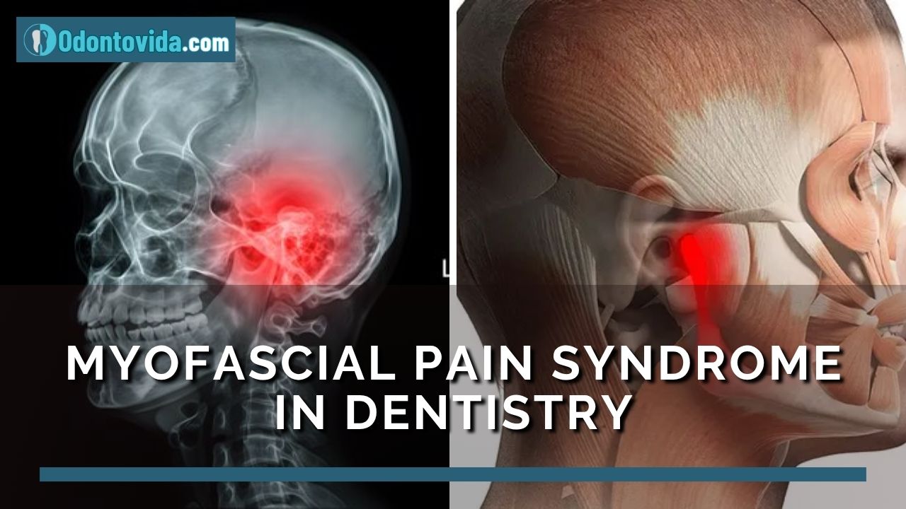
Myofascial Pain Syndrome in Dentistry: Clinical Impact and Modern Management

Myofascial Pain Syndrome

Orofacial pain is a common concern in dental practice and may stem from various sources including dental, joint, neuropathic, or muscular origins.

📌 Recommended Article :
PD/Video 🔽 Bruxism: What are the symptoms? How is it diagnosed? ... Bruxism can occur at any age, and must be diagnosed and treated in time to avoid its serious consequences and complications. Treatment is sometimes usually multidisciplinary
Myofascial pain syndrome (MPS) is one of the most prevalent muscular causes of orofacial and jaw pain, often presenting with facial trigger points and mimicking temporomandibular joint dysfunction (TMJ disorder) or tooth pain. Early recognition is key to avoiding misdiagnosis and unnecessary dental procedures.

Advertisement

Definition of Myofascial Pain Syndrome
MPS is defined as a chronic pain condition involving myofascial trigger points—hyperirritable spots located within taut bands of skeletal muscle or fascia. When palpated, these points produce localized or referred pain and muscular stiffness (Simons et al., 1999; Gerwin, 2020).

Causes and Risk Factors
Several factors contribute to the onset of MPS, including:

° Bruxism and chronic jaw clenching
° Muscle overuse (e.g., prolonged chewing, poor posture)
° Emotional stress, leading to muscle tension
° Poor occlusion or dental misalignment
° Tooth loss or unbalanced prosthetics
° Jaw trauma or repetitive microtrauma

These triggers can result in dysfunctional muscle contraction and sustained activation of trigger points.

📌 Recommended Article :
Video 🔽 Dislocation ... Dislocation of the temporomandibular joint is a painful condition that occurs when the mandibular condyle becomes fixed in the anterosuperior aspect of the articular eminence
Symptoms and Clinical Presentation
Typical symptoms of MPS in dental patients include:

° Persistent jaw pain or soreness
° Facial muscle stiffness and tenderness
° Referred pain to the teeth, temples, ears, or neck
° Jaw fatigue or tightness during talking or chewing
° Clicking or limited range of motion in the TMJ
° Sensation of malocclusion without clinical evidence

These symptoms often resemble TMD or neuropathic conditions, making clinical evaluation essential.

Clinical Relevance in Dentistry
Myofascial pain can complicate dental diagnosis and management due to symptom overlap with:

° Atypical toothache (non-odontogenic pain)
° Temporomandibular joint disorders (TMJ/TMD)
° Facial nerve pain or neuralgia
° Persistent post-treatment dental pain

According to the American Academy of Orofacial Pain (2022), up to 85% of patients with chronic orofacial pain have a muscular component. Failing to recognize MPS can result in unnecessary root canals, extractions, or surgical interventions.

📌 Recommended Article :
Video 🔽 What are the symptoms of clenched teeth and how can we fix it? ... This activity is nocturnal and in states of unconsciousness. The consequences of bruxism is varied, since it affects teeth, muscles and bones
Diagnosis
MPS diagnosis is clinical and based on:

° Palpation of active trigger points
° Reproduction of referred pain
° Muscle tightness and tenderness
° Exclusion of dental or joint pathologies

Imaging is not typically required but may be helpful in ruling out other causes. Ultrasound and electromyography are sometimes used for muscle assessment.

Treatment Options
Effective management of MPS is multidisciplinary and includes:

➤ Physical Therapy and Manual Techniques
°Myofascial release and massage therapy
° Dry needling
° Jaw stretching and strengthening exercises
° Ultrasound therapy or heat application

➤ Medications
° Nonsteroidal anti-inflammatory drugs (NSAIDs)
° Muscle relaxants (e.g., cyclobenzaprine)
° Local anesthetic injections for trigger point relief

➤ Dental Management
° Occlusal adjustments and bite correction
° Night guards or splint therapy for bruxism
° Replacement of missing teeth to restore occlusal balance

➤ Psychological and Behavioral Support
° Cognitive-behavioral therapy for stress and anxiety
° Biofeedback or relaxation techniques

📌 Recommended Article :
Video 🔽 Massage Tutorial: Myofascial release for TMJ/jaw pain ... The temporomandibular joint can undergo some alterations that cause pain to the patient, and can limit the movements of the jaw. In this syndrome the muscles are also involved
💡 Conclusion
Myofascial pain syndrome is a common yet frequently overlooked source of jaw muscle pain and orofacial dysfunction in dental patients. Its overlapping symptoms with other dental and TMJ conditions can lead to misdiagnosis and overtreatment. Dentists must be equipped to recognize the signs of MPS and apply integrated approaches involving manual therapy, medication, and occlusal management. Early diagnosis is critical for successful pain relief and restoration of normal function.

📚 References

✔ American Academy of Orofacial Pain. (2022). Orofacial Pain: Guidelines for Assessment, Diagnosis, and Management (6th ed.). Quintessence Publishing.

✔ Gerwin, R. D. (2020). Classification, epidemiology, and etiology of myofascial pain syndrome. Current Pain and Headache Reports, 24(5), 1–6. https://doi.org/10.1007/s11916-020-00832-5

✔ Simons, D. G., Travell, J. G., & Simons, L. S. (1999). Myofascial Pain and Dysfunction: The Trigger Point Manual (2nd ed.). Williams & Wilkins.

✔ Fernández-de-Las-Peñas, C., & Dommerholt, J. (2018). Myofascial Trigger Points: Pathophysiology and Evidence-Informed Diagnosis and Management. Jones & Bartlett Learning.

✔ Manfredini, D., & Guarda-Nardini, L. (2020). Myofascial pain in temporomandibular disorders: An updated review on diagnosis and management. Journal of Oral Rehabilitation, 47(5), 670–682. https://doi.org/10.1111/joor.12930

📌 More Recommended Items

What is temporomandibular disorder? - Concept, diagnosis and current treatment
Is there a relationship between headache and temporomandibular disorder?
Can children suffer from bruxism?

miércoles, 2 de julio de 2025

Acetaminophen for Kids: Safe Pain Relief in Pediatric Dental Patients

Acetaminophen

Effective pain control is essential in pediatric dentistry to promote positive dental experiences and reduce anxiety.

📌 Recommended Article :
PDF 🔽 Antibiotics and its use in pediatric dentistry: A review ... Antibiotics are commonly used in dentistry for prophylactic as well as for therapeutic purposes. Very often antibiotics are used in unwarranted situations, which may give rise to resistant bacterial strains
Among over-the-counter analgesics, acetaminophen (paracetamol) is widely regarded as a safe and effective option for managing mild to moderate dental pain in children. Its favorable safety profile and accessibility make it a cornerstone in everyday pediatric dental care in the United States.

Advertisement

Mechanism of Action
Acetaminophen primarily works by inhibiting the cyclooxygenase (COX) enzymes in the central nervous system, particularly COX-3. This reduces the production of prostaglandins, resulting in analgesic and antipyretic effects. Unlike nonsteroidal anti-inflammatory drugs (NSAIDs), acetaminophen has minimal anti-inflammatory activity, making it suitable when pain relief is needed without gastrointestinal side effects.

📌 Recommended Article :
PDF 🔽 Guideline on use of antibiotic therapy for pediatric dental patients ... However, the widespread use of antibiotics has permitted common bacteria to develop resistance to drugs that once controlled them
Recommended Pediatric Dosage
According to the American Academy of Pediatrics (AAP) and the U.S. Food and Drug Administration (FDA), the following pediatric dosage guidelines apply:

➤ Oral Dosage for Children:
° 10–15 mg/kg per dose every 4 to 6 hours as needed.
° Maximum Daily Dose: 75 mg/kg/day or no more than 4,000 mg/day (whichever is lower).
➤ Common Forms in the U.S.:
° Infant drops (160 mg/5 mL)
° Children's syrup (160 mg/5 mL)
° Chewable tablets (usually 80 mg or 160 mg)
° Suppositories (vary by age and weight)
Example: A child weighing 44 lbs (20 kg) can receive 200–300 mg per dose every 6 hours, with a maximum of 1,200 mg in 24 hours.

📌 Recommended Article :
PDF 🔽 Pharmacological treatment of oral infections in pediatric dentistry - Recommendations and dose calculation ... In comparison, the metabolism of a drug in a child is different from that of an adult, so the administration of an antibiotic should be taking into account the diagnosis, age and weight of the pediatric patient
Contraindications and Warnings
While acetaminophen is generally safe, there are important situations where its use must be carefully considered:

➤ Absolute Contraindications:
° Known allergy or hypersensitivity to acetaminophen
° Severe liver disease or hepatic failure
➤ Caution in the Following Cases:
° Chronic malnutrition or dehydration
° Use in neonates (requires adjusted dosing and close monitoring)
° Accidental overdose due to combination with other OTC medications containing acetaminophen
Important: Caregivers should be educated to avoid combining multiple products (e.g., cold medications) that may contain acetaminophen.

📌 Recommended Article :
PDF 🔽 Dental pain in pediatric dentistry - Assessment tools ... We share an article that reviews the important aspects of pain in pediatric dentistry, and the tools for a correct evaluation today
Clinical Evidence in Pediatric Dentistry
Recent studies support acetaminophen’s effectiveness in managing dental pain in children, especially following common procedures such as extractions, pulp therapy, or trauma management.
A clinical trial by Coelho et al. (2021) found that acetaminophen provided pain relief equivalent to ibuprofen after dental procedures in children, with fewer gastrointestinal side effects. The American Academy of Pediatric Dentistry (AAPD, 2023) also endorses acetaminophen as the first-line analgesic for young children or those who cannot take NSAIDs.

📌 Recommended Article :
Video 🔽 How To Determine a Child's Medication Dose ... All the necessary data should be taken to be able to calculate the dose in children. Many take the adult dose as a reference, but this could lead to problems such as poisoning or drug resistance
💬 Discussion Acetaminophen remains a go-to option in pediatric dental care due to its strong safety profile and efficacy. Weight-based dosing is essential, and dental professionals must provide clear instructions to caregivers to prevent misuse or overdose. In some cases of moderate to severe inflammation, NSAIDs may offer superior pain control, but acetaminophen is often preferred in children due to fewer side effects.
Combination therapy (e.g., acetaminophen plus ibuprofen) may be considered in select cases under dental supervision.

💡 Conclusion
Acetaminophen is a safe and effective pain reliever for pediatric dental patients when used at the correct dosage and with proper caregiver guidance. Understanding its mechanism, indications, and safety limits ensures optimal pain management and prevents complications related to improper use.

📚 References

✔ American Academy of Pediatrics. (2023). Pain Management Guidelines for Pediatric Patients. Retrieved from https://www.aap.org

✔ American Academy of Pediatric Dentistry. (2023). Use of Analgesics in Pediatric Dental Care. Retrieved from https://www.aapd.org

✔ U.S. Food and Drug Administration (FDA). (2023). Acetaminophen and Safe Use in Children. Retrieved from https://www.fda.gov

✔ Coelho, M. S., Oliveira, D., & Silva, A. C. (2021). Comparative effectiveness of paracetamol and ibuprofen for post-operative pain in pediatric dental patients. Pediatric Dentistry, 43(1), 45–50.

✔ World Health Organization. (2023). WHO Model List of Essential Medicines for Children – 8th Edition. Retrieved from https://www.who.int

📌 More Recommended Items

Pain and anxiety management for pediatric dental procedures using various combinations of sedative drugs: A review
What are the medicines used in pediatric dentistry?
Appropriate use of antibiotics in pediatric odontogenic infections

martes, 1 de julio de 2025

Effective Pediatric Oral Surgery Management: Proven Strategies for a Positive Experience

Oral Surgery

Oral surgery in children—such as extractions, frenectomies, and removal of supernumerary teeth—presents unique challenges. These include dental anxiety, limited communication abilities, and emotional sensitivity.

📌 Recommended Article :
Video 🔽 How To Prevent A Dry Socket ... At the time that the surgical procedure is completed, the surgeon must instruct the patient on the care that must be taken to avoid an infectious process
A successful surgical experience depends on more than clinical technique; it also relies on emotional support, behavior management, and a child-friendly environment. Dental providers must apply comprehensive strategies that meet the developmental, emotional, and psychological needs of young patients.

Advertisement

Pediatric Patient Characteristics in Oral Surgery
Children differ significantly from adults in their response to clinical settings:

° Cognitive development affects their understanding of dental procedures.
° Emotional response includes fear of pain, separation anxiety, and past traumatic experiences.
° Communication skills are often limited, especially in younger children.
° Recognizing these traits is critical in developing patient-centered care strategies.

📌 Recommended Article :
PDF 🔽 Guideline on Pediatric Oral Surgery ... Management of children under sedation or general anesthesia requires extensive training and expertise
Strategies for a Positive Pediatric Oral Surgery Experience

1. Comprehensive Preoperative Assessment
Key steps include:

° Assessing dental anxiety using tools like the Venham Picture Test or FLACC scale.
° Reviewing prior dental experiences and behavior patterns.
° Providing child-friendly explanations with visual aids and interactive models.

2. Behavior Management Techniques
Recommended by the American Academy of Pediatric Dentistry (AAPD):

° Tell-Show-Do: Builds trust and predictability.
° Positive reinforcement: Verbal praise, stickers, or small rewards.
° Voice control: Calm but firm tone to guide behavior.
° Distraction: Videos, music, or storytelling to divert attention.
° Parental presence: Used selectively based on the child's needs and behavior.

📌 Recommended Article :
Video 🔽 How to control bleeding after extraction? ... Post-treatment bleeding is a common complication after tooth extraction, so it is important that the dentist makes recommendations to avoid any type of problems
3. Pharmacologic Management
Used when non-pharmacological techniques are insufficient:

° Nitrous oxide sedation: Safe and widely used for mild to moderate anxiety.
° Oral sedation: Midazolam and similar agents for more significant anxiety.
° General anesthesia: Reserved for extensive procedures or severe dental phobia, typically in a hospital setting.

4. Child-Friendly Dental Environment
Environmental modifications include:

° Soft colors and playful decor in the operatory.
° Dental tools with friendly designs or names.
° Friendly, trained staff skilled in pediatric communication.

📌 Recommended Article :
Dental Article 🔽 8 interesting facts about the Wisdom Tooth ... While they often emerge in late adolescence or early adulthood, their presence and impact on oral health have been subjects of extensive study
5. Effective Communication with Children and Parents
Strong communication builds trust and improves cooperation:

° Clear and simple explanations tailored to the child’s developmental level.
° Transparent discussion of surgical procedures and aftercare with parents.
° Giving the child limited choices (e.g., music selection) to increase their sense of control.

💬 Discussion
Research shows that untreated dental fear in childhood often continues into adulthood, negatively affecting oral health outcomes (Klingberg & Broberg, 2022). Combining behavior management with appropriate sedation techniques significantly reduces anxiety and improves surgical cooperation (Olumide et al., 2021). A child-focused, holistic approach is increasingly recognized as the gold standard in pediatric oral surgery, emphasizing both technical excellence and emotional support.

📌 Recommended Article :
PDF 🔽 Pulpotomy Medicaments used in Deciduous Dentition: An Update ... It is important that all clinicians, particularly for pediatric dentists, be up to date with the recent trends in this area of dental treatment for children
💡 Conclusions
Successful pediatric oral surgery involves more than technical skill. It requires tailored strategies that consider each child’s emotional, psychological, and developmental needs. The integration of behavior management, pharmacological tools, environmental modifications, and effective communication fosters a positive experience that encourages lifelong oral health habits.

📚 References

✔ American Academy of Pediatric Dentistry. (2023). Behavior Guidance for the Pediatric Dental Patient. The Reference Manual of Pediatric Dentistry. https://www.aapd.org/globalassets/media/policies_guidelines/bp_behavior.pdf

✔ Klingberg, G., & Broberg, A. G. (2022). Dental fear/anxiety and dental behaviour management problems in children and adolescents: A review of prevalence and concomitant psychological factors. International Journal of Paediatric Dentistry, 32(1), 3–9. https://doi.org/10.1111/ipd.12898

✔ Olumide, F., Costa, L. R., & Almeida, E. S. (2021). Behavioral techniques versus pharmacological management in pediatric dental sedation: A systematic review. Pediatric Dentistry, 43(2), 97–105. https://www.aapd.org/research/oral-health-policies--recommendations/behavior-guidance/

✔ Versloot, J., Veerkamp, J. S. J., & Hoogstraten, J. (2020). Children’s coping with pain during dental care. Community Dentistry and Oral Epidemiology, 48(4), 272–278. https://doi.org/10.1111/cdoe.12534

📌 More Recommended Items

Mandibular tumors in pediatric patients. Report of 04 cases of aggressive tumors
Marsupialization of a large mandibular cyst in a pediatric patient - Clinical Case
What is frenectomy? Step by step surgical procedure

Pulp Polyps in Children: Causes, Diagnosis, and Treatment Options in Pediatric Dentistry

Pulp Polyps

Chronic hyperplastic pulpitis, commonly referred to as a pulp polyp, is a non-neoplastic, benign proliferation of pulpal tissue. It is often observed in children and adolescents, attributed to their rich pulpal vascularity and strong immune response.

📌 Recommended Article :
PDF 🔽 Pulpotomy Medicaments used in Deciduous Dentition: An Update ... It is important that all clinicians, particularly for pediatric dentists, be up to date with the recent trends in this area of dental treatment for children
While it is a protective response to chronic inflammation, it reflects significant pulpal exposure, often requiring endodontic or surgical management.

Advertisement

Etiology
Pulp polyps arise primarily due to:

➤ Advanced Dental Caries: The most common cause, where prolonged exposure leads to bacterial infiltration and chronic inflammation (Seltzer & Bender, 2002).
➤ Dental Trauma: Fractured teeth or open pulp chambers expose the tissue to irritation.
➤ Open Apex or Immature Roots: In children, an open apex allows for vascular proliferation and granulation tissue growth (Brito et al., 2018).
➤ Poor Oral Hygiene: Facilitates microbial colonization and chronic irritation.

📌 Recommended Article :
Video 🔽 What is the difference between apexogenesis and apexification? ... In the presence of an open apex, apexogenesis (apicoformation) or apexification can be used. Both treatments have differences that will be resolved with the video that we show below
Clinical Features

° Appears as a red or pink soft tissue mass arising from a large carious lesion or a fractured tooth.
° Painless in most cases due to lack of nerve innervation in the granulation tissue.
° No bleeding on manipulation or mild bleeding only.
° Most commonly seen in molars of children and adolescents.
° May interfere with occlusion or become traumatized during chewing.

📌 Recommended Article :
Video🔽 Dental Treatment: Vital Pulp Therapy for Primary Teeth ... The anatomical characteristics of primary teeth are different from permanent teeth. It is important to know these differences when performing any dental procedure
Diagnosis
Diagnosis is primarily clinical, but may be supported by:

➤ Radiographs: To evaluate the extent of decay and assess root development or periapical pathology.
➤ Pulp Vitality Testing: Usually not necessary, but may help in borderline cases.
➤ Histopathology: Shows fibrovascular granulation tissue with chronic inflammatory cells (Ricucci & Siqueira, 2010).

📌 Recommended Article :
Video 🔽 Apexogenesis: Step by step procedure ... We share a video that explains everything about apexogenesis: definition, indications, contraindications and the step-by-step procedure
Differential Diagnosis

° Gingival polyps
° Papillary hyperplasia
° Peripheral giant cell granuloma
° Inflammatory fibrous hyperplasia

📌 Recommended Article :
Video 🔽 Pulpotomy: Diagnosis, types and procedure ... During the procedure, the caries and the dental pulp are partially removed, thus maintaining the vitality and permanence of the primary tooth in the mouth
Treatment Options
Treatment depends on the tooth’s vitality, restorability, and root development stage:

1. Pulpotomy
Indicated in vital teeth with no periapical pathology. The coronal pulp is removed and the radicular pulp preserved, often using materials like MTA or Biodentine (El Meligy et al., 2019).
2. Pulpectomy
Complete removal of pulp tissue, ideal for non-vital primary teeth. Root canals are filled with resorbable materials like ZOE or iodoform-based pastes.
3. Extraction
Indicated in non-restorable teeth, teeth with excessive root resorption, or when endodontic treatment is contraindicated. Placement of a space maintainer may be necessary.

📌 Recommended Article :
PDF 🔽 Manual of diagnosis and pulp treatment in non-vital primary teeth ... A correct history, clinical and radiographic evaluation is necessary in these cases to determine the appropriate treatment for these cases (dental extraction, pulpectomy, lesion sterilization tissue repair)
Prognosis and Follow-Up
With appropriate intervention, the prognosis is excellent. Follow-up is essential to monitor eruption of permanent teeth and prevent space loss.

💬 Discussion
Pulp polyps are often misunderstood as aggressive lesions, but they are a benign response to chronic irritation in young, healthy pulp tissue. Pediatric patients’ immune and reparative capacities allow this type of response, unlike in adults. Early detection through routine exams can prevent complications. Advances in biocompatible materials and minimally invasive dentistry have improved outcomes for pulp therapy in children.

💡 Conclusion
Pulp polyps in children are a clear indicator of neglected dental caries or trauma. Prompt recognition and appropriate treatment—whether pulp therapy or extraction—are critical for preserving oral health and preventing long-term complications in developing dentition.

📚 References

✔ Brito, F. C., de Sousa, C. M., & Maia, L. C. (2018). Pediatric endodontic treatment: A systematic review. International Journal of Paediatric Dentistry, 28(6), 525–539. https://doi.org/10.1111/ipd.12406

✔ El Meligy, O. A., Allazzam, S. M., & Alamoudi, N. M. (2019). Clinical and radiographic success of MTA pulpotomy in primary molars: A 24-month follow-up. European Archives of Paediatric Dentistry, 20(3), 225–230. https://doi.org/10.1007/s40368-019-00414-3

✔ Ricucci, D., & Siqueira, J. F. (2010). Biofilms and apical periodontitis: Study of prevalence and association with clinical and histopathologic findings. Journal of Endodontics, 36(8), 1277–1288. https://doi.org/10.1016/j.joen.2010.04.007

✔ Seltzer, S., & Bender, I. B. (2002). The dental pulp: Biology, pathology, and regenerative therapies. Quintessence Publishing.

📌 More Recommended Items

Root Canal Treatment Procedure - Step by step
Management of Endodontic Emergencies: Pulpotomy Versus Pulpectomy
Technique for Primary Molar Tooth Pulpotomy

domingo, 29 de junio de 2025

Top Benefits of Chemical Caries Removal in Children and Patients with Dental Anxiety

Chemical Caries Removal

Modern dentistry increasingly embraces minimally invasive approaches that prioritize preserving healthy tooth structure and enhancing the patient experience.

📌 Recommended Article :
Video🔽 Use of Silver Diamine Fluoride (SDF) - General Guide on its application ... It offers several advantages such as its easy application, low cost, it avoids the use of dental anesthetics, and it can be applied in places that do not have dental equipment
In this context, chemical caries removal (CCR) has emerged as a safe, effective alternative to traditional drilling—especially valuable in pediatric dentistry and for patients with dental anxiety or phobia. This technique allows clinicians to eliminate decayed dentin without rotary tools, reducing discomfort, fear, and the need for local anesthesia.

Advertisement

What Is Chemical Caries Removal?
CCR is a conservative, non-invasive technique that uses enzymatic or oxidizing agents to soften infected dentin, allowing its manual removal without mechanical drilling. Common products include Carisolv®, Papacárie Duo®, Brix3000®, and Carie-Care™, which have proven effective in clinical studies.

Why Is CCR Ideal for Children and Anxious Patients?

1. Avoids the dental drill
The sound and vibration of a dental drill often trigger anxiety in children and phobic adults. CCR eliminates the need for rotary instruments.
2. Often requires no local anesthesia
Recent studies show that chemical agents can remove caries painlessly, making injections unnecessary in many cases (Elgalaid et al., 2022).
3. Increases treatment acceptance
Minimally invasive techniques result in a more relaxed experience, improving cooperation in young children and anxious patients (Ghasempour et al., 2020).
4. Preserves healthy tooth structure
Most CCR products act selectively on infected dentin, aligning with the principles of minimally invasive dentistry.
5. Creates a calm clinical environment
Less noise and vibration help maintain a soothing atmosphere, reducing stress for both patient and provider.

📌 Recommended Article :
PDF 🔽 Molar incisor hypomineralisation: Definition, diagnosis and clinical management ... In comparison, the metabolism of a drug in a child is different from that of an adult, so the administration of an antibiotic should be taking into account the diagnosis, age and weight of the pediatric patient
Latest Scientific Evidence
Recent clinical research highlights CCR’s effectiveness and patient acceptance:

° Elgalaid et al. (2022) found that Carisolv® significantly lowered anxiety and pain perception in children compared to conventional methods.
° Ghasempour et al. (2020) reported high satisfaction rates using Brix3000® among children aged 4–7 years.
° Santos et al. (2021) confirmed the safety and effectiveness of Papacárie Duo® in patients with mild to moderate dental anxiety.

📌 Recommended Article :
PDF 🔽 Alternative for the rehabilitation of pediatric patients with early childhood caries ... We share the oral rehabilitation of a pediatric patient affected by early childhood caries, using a functional appliance aesthetically appropriate for the patient
Clinical Guidelines

° Indications: Ideal for shallow to moderate dentin caries without pulpal involvement.
° Contraindications: Avoid in deep lesions near the pulp or sclerotic dentin.
° Chair time: Slightly longer than conventional drilling, but often compensated by improved cooperation.
° Restoration options: Compatible with adhesive and bioactive restorative materials.

📌 Recommended Article :
PDF 🔽 Pharmacological treatment of oral infections in pediatric dentistry - Recommendations and dose calculation ... In comparison, the metabolism of a drug in a child is different from that of an adult, so the administration of an antibiotic should be taking into account the diagnosis, age and weight of the pediatric patient
💬 Discussion
Chemical caries removal represents a paradigm shift in managing caries in vulnerable populations. Its patient-friendly and drill-free nature allows for a less traumatic and more empathetic experience. The ability to reduce fear and discomfort during treatment makes CCR especially valuable in pediatric and behavioral dentistry. It also fosters better long-term dental relationships by building trust from an early age.

💡 Conclusion
Chemical caries removal is a powerful tool for modern dentistry, particularly when treating children and patients with dental phobia. Backed by recent evidence, its use promotes a more comfortable, conservative, and effective dental care experience. CCR should be considered a standard part of the clinical toolkit when aiming for anxiety-free dental visits.

📚 References

✔ Elgalaid, M. A., Alshoraim, M. A., Alhazmi, Y. F., & Alahmari, R. A. (2022). A randomized clinical trial comparing Carisolv and rotary instruments in caries removal: anxiety and pain perception in pediatric patients. BMC Oral Health, 22, 333. https://doi.org/10.1186/s12903-022-02458-4

✔ Ghasempour, M., Yeganeh, P., & Golkari, A. (2020). Comparison of the effectiveness of Brix3000 and conventional methods in caries removal in children. Journal of Dentistry for Children, 87(3), 151–156.

✔ Santos, A. P., Freire, M. C. M., Oliveira, B. H., & Paiva, S. M. (2021). Effectiveness of Papacárie Duo in minimally invasive treatment of dental caries in anxious children: a randomized clinical trial. Pediatric Dentistry, 43(4), 259–265.

✔ Lussi, A., & Schaffner, M. (2019). Advances in minimally invasive caries removal: Chemo-mechanical agents. In Mount, G. J. & Hume, W. R. (Eds.), Preservation and Restoration of Tooth Structure (3rd ed., pp. 97–104). Wiley-Blackwell.

📌 More Recommended Items

Chemical Caries Removal: Drill-Free Technique, Materials, and Clinical Application
Glass Ionomer Cement in Pediatric Dentistry: Benefits, Uses, and Step-by-Step Application Guide
How well do fluoride treatments work at preventing tooth decay?