Mostrando entradas con la etiqueta Oral Medicine. Mostrar todas las entradas

jueves, 1 de diciembre de 2022

Molar incisor hypomineralisation: Definition, diagnosis and clinical management

Molar incisor hypomineralisation

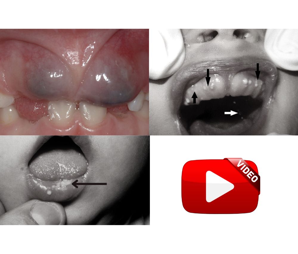
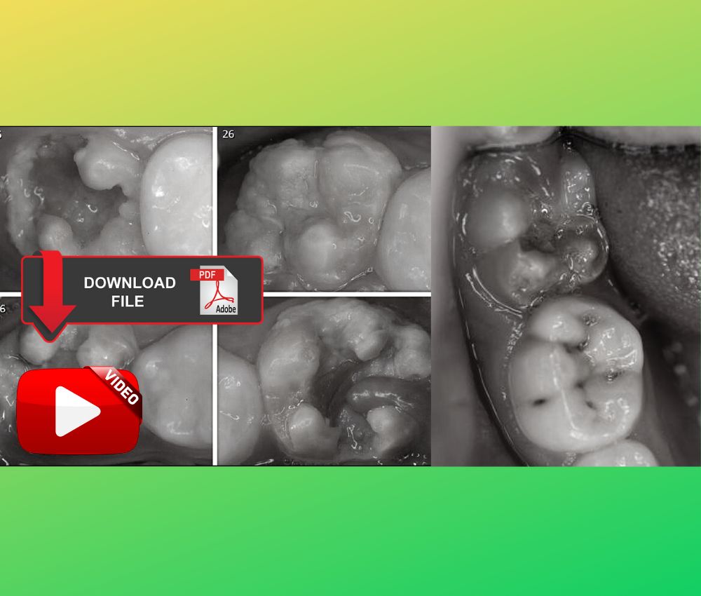
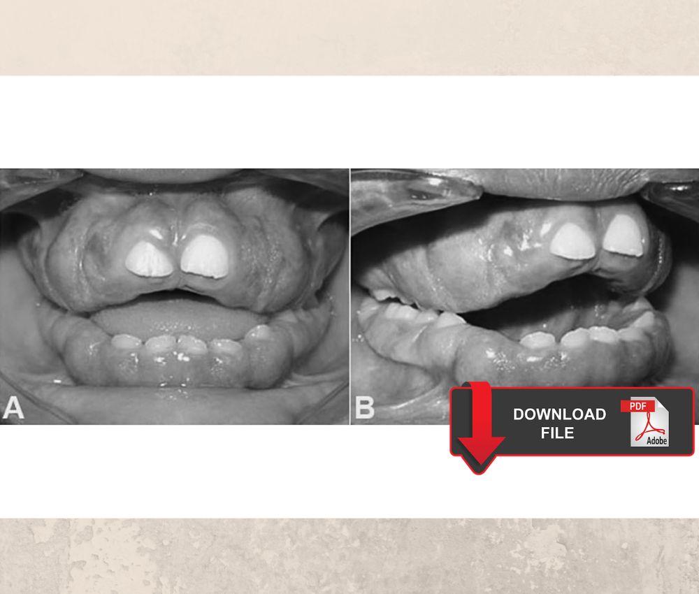
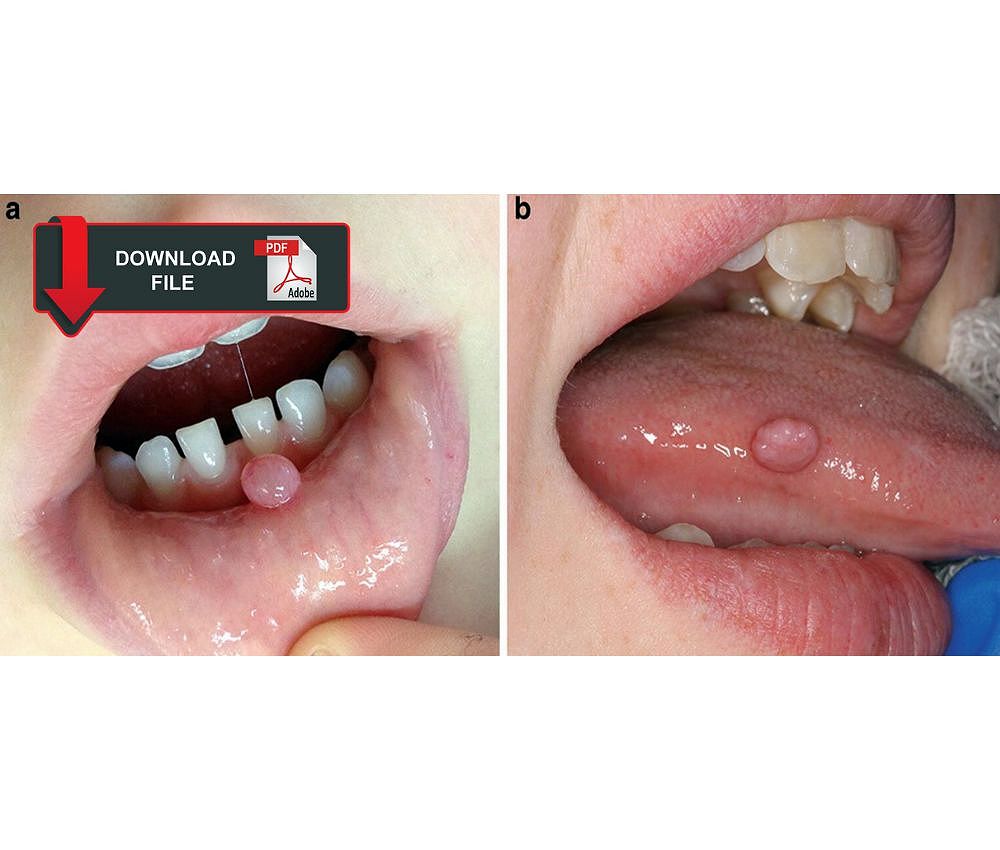
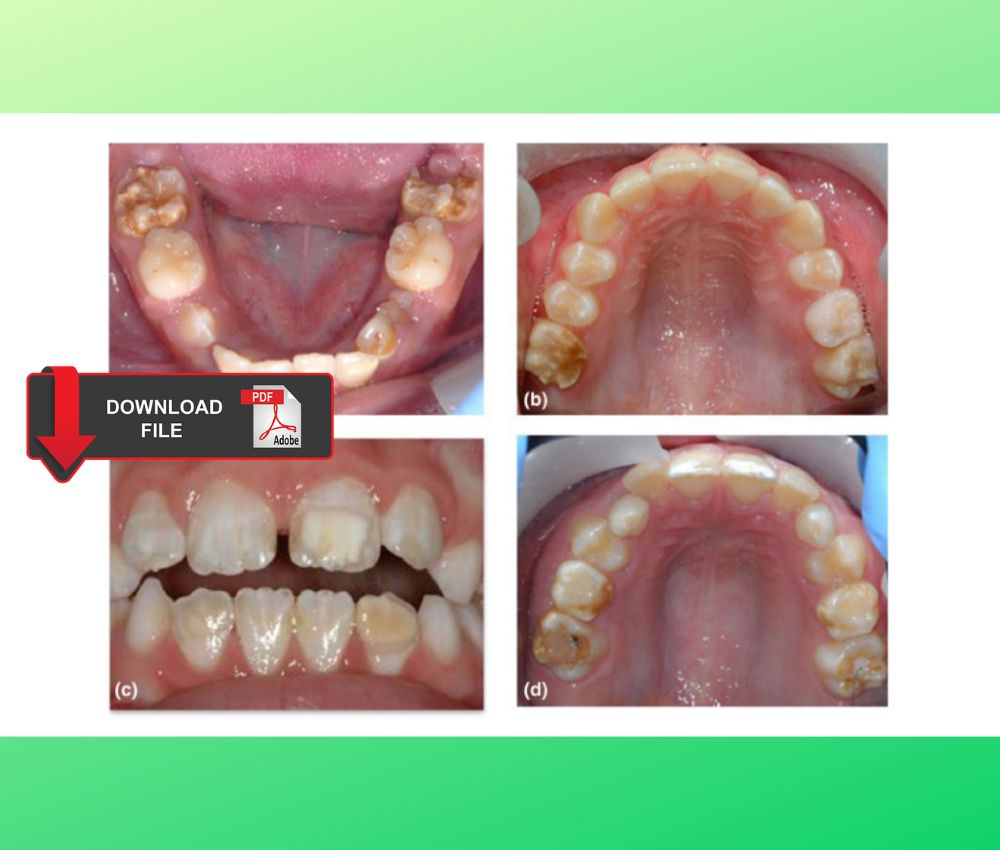
Molar incisor hypomineralization is a development defect of dental enamel, and affects the aesthetics of the tooth and generates sensitivity and a high degree of enamel fracture, in addition to a high risk of caries.

📌Recommended Article :
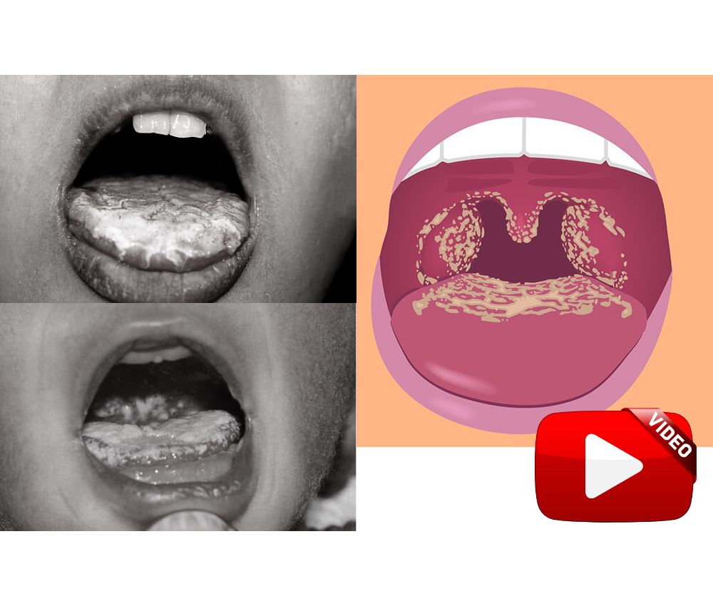
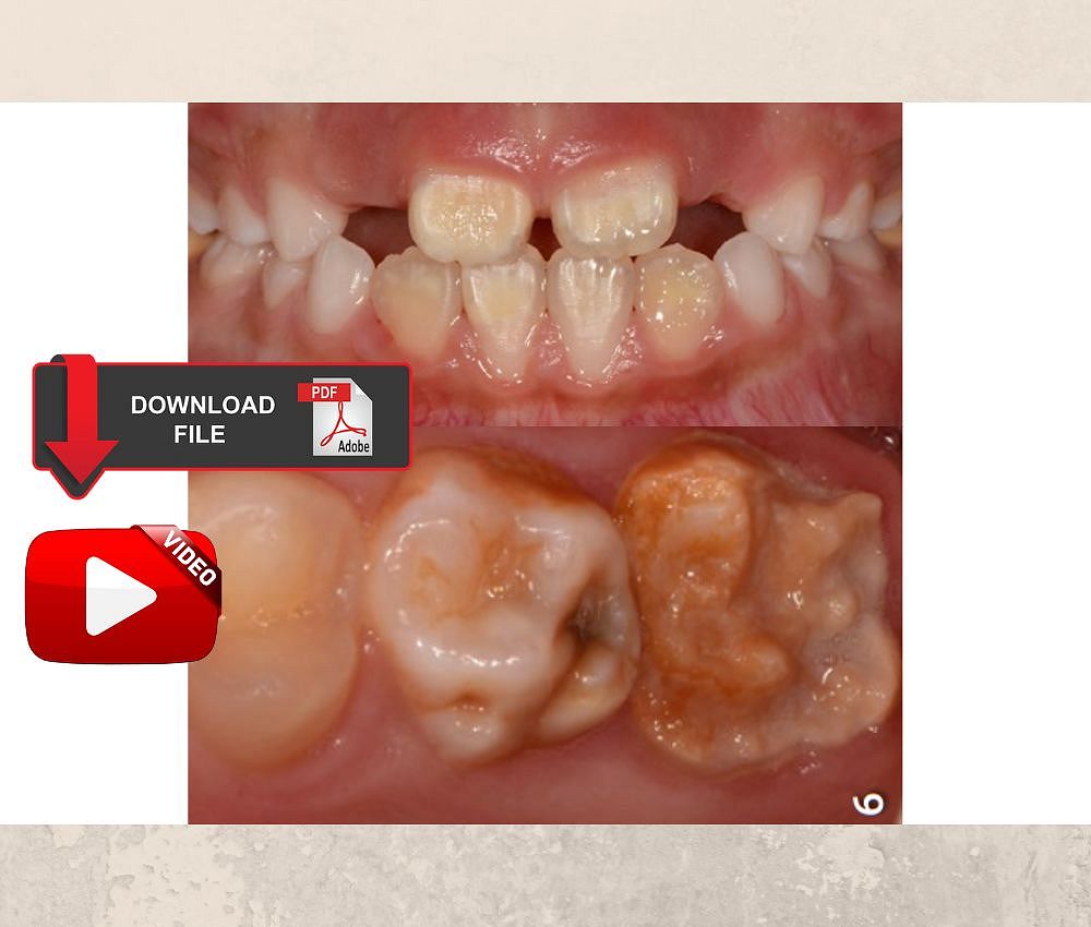
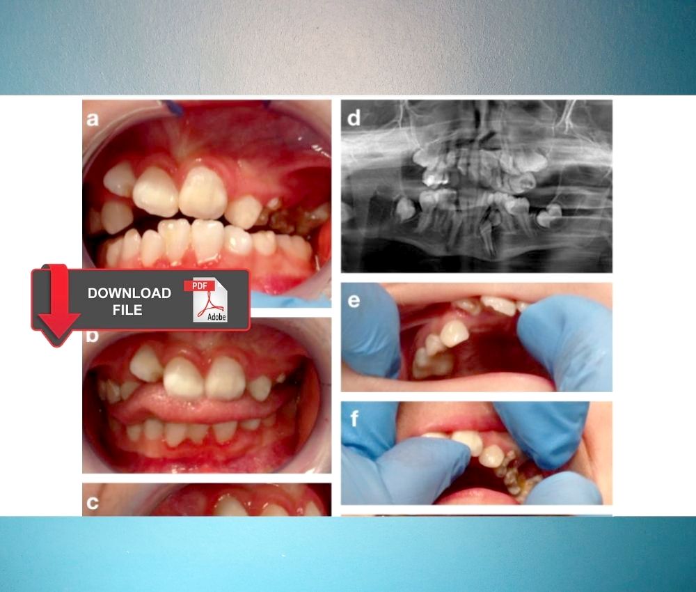
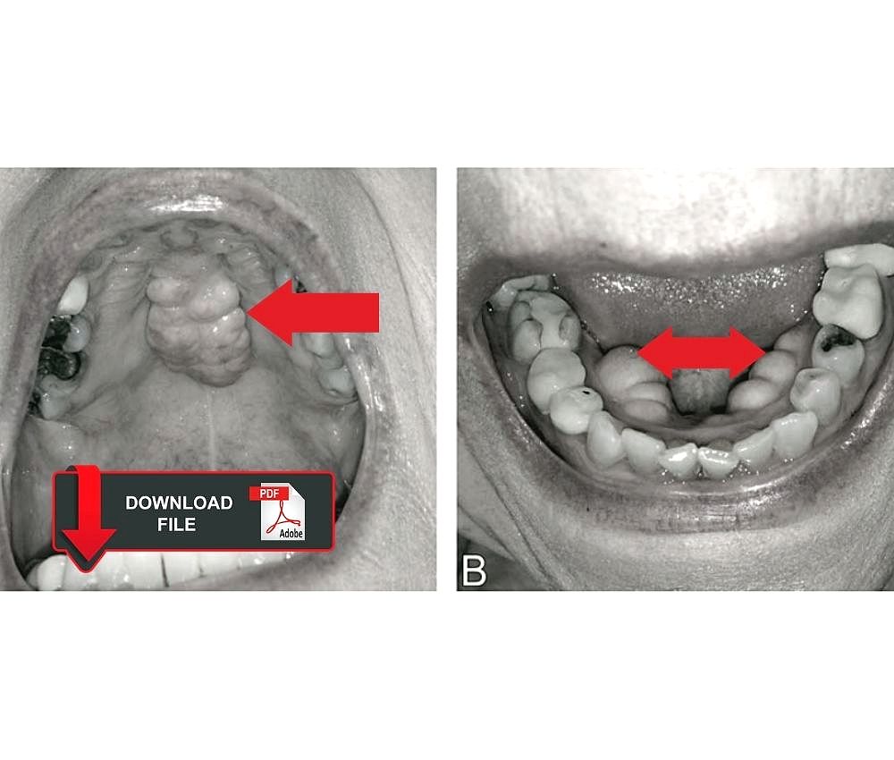
Download the PDF 🔽 Molar Incisor Hypomineralization: Minimally Invasive Treatments - Step by Step ... The tooth affected by molar incisor hypomineralization has a porous appearance with brown, yellow and opaque white stains

The molar or incisor affected by hypomineralization presents a white/yellow color, and with an enamel susceptible to fracture. There are mild and severe cases, which is why an early diagnosis is important to avoid tooth loss.

Advertisement

We share an article that teaches us what is the clinical management of molar incisor hypomineralization (MIH), differential diagnosis and treatment in mild and severe cases in children.

📌Download the article in PDF :

👉 READ AND DOWNLOAD "Molar incisor hypomineralisation: Definition, diagnosis and clinical management" IN FULL IN PDF👈


Daly D, Waldron JM. Molar incisor hypomineralisation: clinical management of the young patient. J Ir Dent Assoc. 2009 Apr-May;55(2):83-6. PMID: 19455847.

📌 More Recommended Items

ORAL REHABILITATION of a child with dentinogenesis imperfecta
Molar-Incisor Hypomineralisation: Clinical Manifestations and Treatment
What is tooth sensitivity?

lunes, 21 de noviembre de 2022

How to treat anaphylaxis in children? - Clinical manifestations and treatment

anaphylaxis

Anaphylaxis is a severe and life-threatening allergic reaction in response to an allergen. The response is diverse and can range from skin manifestations to cardiovascular or respiratory disorders.

📌Recommended Article :
Download the PDF 🔽 Allergic manifestations to local anaesthetic in pediatric dentistry: Prevention and management ... Allergic reactions to local anesthetics in pediatric patients are not frequent, but we must take the necessary preventive measures and know how to manage hypersensitivity to anesthetics.

The clinical picture must be recognized in order to take immediate action and avoid serious consequences. In dentistry, a picture of anaphylaxis may occur due to contact with a drug or an allergenic agent such as latex.

Advertisement

We share important information about the clinical picture of anaphylaxis, the preventive attitude and the appropriate treatment for a pediatric patient in dentistry.

📌Download the article in PDF :

👉 READ AND DOWNLOAD "How to treat anaphylaxis in children? - Clinical manifestations and treatment" IN FULL IN PDF👈


Cheng A. Emergency treatment of anaphylaxis in infants and children. Paediatr Child Health. 2011 Jan;16(1):35-40. PMID: 22211074; PMCID: PMC3043023.

📌 More Recommended Items

Local anesthesia calculations: How to avoid overdose and toxicity in pediatric patients?
Considerations for Pediatric Local Anesthesia
Local Anesthetics Allergies in Children

miércoles, 9 de noviembre de 2022

Frequent oral pathologies in the newborn - Diagnosis and treatment (cysts, infections, tumors)

oral-pathology

The newborn can present a series of pathologies that must be detected in time for early treatment. Sometimes the obstetrician or pediatrician does not recognize oral alterations, so evaluation by a pediatric dentist is important.

📌 Recommended Article:
Download the PDF 🔽 Neonatal tooth with Riga-Fede disease affecting breastfeeding: A case report ... Neonatal teeth generate an ulcer in the sublingual region, and it is known as Riga-Fede disease. Breastfeeding is compromised by the discomfort generated by the ulcer.

Some of the pathologies can cause limitations in feeding and in the development of the baby, so it is necessary to explain and guide the parents about the alterations found, the repercussions and the appropriate treatment.

Advertisement

The video that we share details the most common oral pathologies in neonates, detailing their etiology, clinical characteristics, diagnosis and treatment.

📌 More recommended items

Guide for the surgical management and oral pathology of the pediatric patient
Craniofacial syndromes and oral anomalies. Characteristics and clinical cases
Surgical excision of mucocele with local anesthesia in an 8-month-old baby

📌 Watch the video "Frequent oral pathologies in the newborn - Diagnosis and treatment (cysts, infections, tumors)"


Youtube/ Dr Mandana Donoghue

jueves, 3 de noviembre de 2022

What is Oral Candidiasis? - Everything you need to know - Symptoms, diagnosis and treatment

Periodoncia

The Candida Albicans fungus, responsible for oral candidiasis, lives in our oral cavity together with other microorganisms but does not generate any pathological alteration.

📌 Recommended Article:
Download the PDF 🔽 Management of Angular Cheilitis in children ... Angular Cheilitis occurs more in children and it is caused by children sensitivity against certain contact agents like toys, foods, sunlight, allergy against medicines, cosmetics, and long term antibiotic treatment.

The disease manifests itself when the balance of the bacterial flora is disturbed or the patient's immunity decreases. Candida albicans most commonly affects infants and older adults, as they often have reduced immunity.

Advertisement

All the information about Oral Candidiasis and its causes, symptoms, diagnosis and treatment in the video that we share below.

📌 More recommended items

Guide for the surgical management and oral pathology of the pediatric patient
Deformity of the tongue in an infant: Riga-Fede disease
How to Manage a Pediatric Patient with Oral Ulcers

📌 Watch the video "ORAL CANDIDIASIS | Diagnosis and Management "


Youtube/ TaughtWell SimplifyEd

lunes, 31 de octubre de 2022

Molar Incisor Hypomineralization: Minimally Invasive Treatments - Step by Step

Periodoncia

The tooth affected by molar incisor hypomineralization has a porous appearance with brown, yellow and opaque white stains. It is of unknown etiology and the patient is affected in aesthetics.

📌Recommended Article :
Download the PDF 🔽 Modified Hall technique in hypomineralized molar ... We share the management of severely hypomineralized molars using the modified Hall technique, indicating its advantages, objectives and the step-by-step process

The patient may present sensitivity, and an accelerated wear of the dental structure. The risk of caries is high and must be treated to prevent tooth loss.

Advertisement

We share the management protocol and techniques of minimally invasive treatments that exist for molar incisor hypomineralization.

📌Download the article in PDF :

👉 READ AND DOWNLOAD "Molar incisor hypomineralization (MIH): conservative treatment management to restore affected teeth" IN FULL IN PDF👈


Fragelli, Camila & Souza, Juliana & Jeremias, Fabiano & Cordeiro, Rita & Santos-Pinto, Lourdes. (2015). Molar incisor hypomineralization (MIH): conservative treatment management to restore affected teeth. Brazilian oral research.

📌 More Recommended Items

Molar-Incisor Hypomineralisation: Clinical Manifestations and Treatment
Hall technique: Complete information for the treatment of carious primary molars
Stainless Steel Crowns: Adaptation and installation - Step by step

📌 Also watch the video : Minimal Interventions to Manage Molar Incisor Hypomineralization


Fuente: Youtube/ Elevate Oral Care

lunes, 10 de octubre de 2022

Hereditary Gingival Fibromatosis in a pediatric patient - Characteristics and treatment

Hereditary Gingival Fibromatosis

Hereditary gingival fibromatosis is a rare, benign, genetic disorder characterized by progressive enlargement of the gingival tissue. It has no symptoms and can be generalized or localized.

The increase in the gum prevents or delays the eruption of primary or permanent teeth, also affects the aesthetics of the patient, generating diastemas and dental malposition. Treatment is surgical removal of the gingival tissue.

Advertisement

We share the clinical case report of a pediatric patient with hereditary gingival fibromatosis, the clinical and histological characteristics, diagnosis and treatment are addressed.

Oral Medicine


👉 READ AND DOWNLOAD "Hereditary Gingival Fibromatosis in a pediatric patient - Characteristics and treatment" IN FULL IN PDF👈


Cunha JLS, Ramos MACDC, Regis DM, Sanchéz-Romero C, de Andrade ME, Bezerra BT, de Albuquerque-Júnior RLC. Generalized hereditary gingival fibromatosis in a child: clinical, histopathological and therapeutic aspects. Autops Case Rep. 2020 Jan 21;10(1):e2020140.

You may also like :
Marsupialization of a large mandibular cyst in a pediatric patient - Clinical Case
What are those bony bumps in my mouth? (Torus)
Ameloblastoma in a pediatric patient - Characteristics, treatment and clinical case

jueves, 29 de septiembre de 2022

Molar-Incisor Hypomineralisation: Clinical Manifestations and Treatment

Molar-Incisor Hypomineralisation

Clinically, Molar Incisor Hypomineralization is characterized by presenting a dental piece with an enamel that fractures easily and of various colors that vary between yellow, brown and white. The causes are unknown but it is related to external and pharmacological factors.

The patient presents sensitivity, pain on chewing, high risk of caries. Other abnormalities such as fluorosis and enamel hypoplasia should be ruled out.

Advertisement

We share a complete article on the factors, clinical manifestations, diagnosis and management of Molar Incisor Hypomineralization.

Oral Medicine


👉 READ AND DOWNLOAD "Atraumatic Restorative Treatment - Step-by-step procedure manual" IN FULL IN PDF👈


Joëlle A.Dulla Hendrik Meyer-Lückel. Molar-incisor hypomineralisation: narrative review on aetiology, epidemiology, diagnostics, and treatment decision. SWISS DENTAL JOURNAL SSO VOL 131 11 P2021


👉 ALSO WATCH THE VIDEO: Webinar "Hypomineralised enamel (MIH) - coming to a child near you" - Dr. David Manton


Fuente: Youtube / GCEuropeProducts

You may also like :
Stainless Steel Crowns: Adaptation and installation - Step by step
Esthetic Options for Anterior Primary Teeth - Characteristics
Indications for Hall technique usage in pediatric dentistry


miércoles, 17 de agosto de 2022

What is temporomandibular disorder? - Concept, diagnosis and current treatment

TMD

The temporomandibular joint is formed by the mandibular and the temporal bone of the skull, which together with muscular structures allow movements such as opening, closing, laterality, protrusion and mandibular retrusion.

The etiology of temporomandibular disorders are complex and varied, and it is a challenge for the evaluation, diagnosis and management of this pathology. Treatment can be conservative or surgical.

Advertisement

Learn about the clinical manifestations, causes and consequences of temporomandibular disorders, in addition to the management and treatment of this pathology.

TMD


👇 READ AND DOWNLOAD THE ARTICLE "Temporomandibular Disorders: Current Concepts and Controversies in Diagnosis and Management" IN PDF 👇


Li, D.T.S.; Leung, Y.Y. Temporomandibular Disorders: Current Concepts and Controversies in Diagnosis and Management. Diagnostics 2021, 11, 459. https://doi.org/10.3390/diagnostics11030459

You may also like :
Temporomandibular disorders (TMD) in children - Clinical examination and treatment
Is there a relationship between headache and temporomandibular disorder?
What causes jaw pain?

miércoles, 10 de agosto de 2022

Ameloblastoma in a pediatric patient - Characteristics, treatment and clinical case

Oral Surgery

Ameloblastoma is an odontogenic tumor, non-malignant but locally aggressive, with high recurrence, and is evidenced as a facial swelling, or sometimes as a radiographic finding.

The patient refers symptoms such as pain, dental mobility, malocclusion and others. Treatment is surgical, and can be conservative or radical, and the technique to be used will depend on the pathological and radiographic evaluation of the tumor.

Advertisement

We share a review on ameloblastoma and its clinical and radiographic characteristics, as well as surgical treatment, regarding the presentation of the case in a pediatric patient.

Oral Surgery


👉 READ AND DOWNLOAD "Ameloblastoma in a pediatric patient - Characteristics, treatment and clinical case" IN FULL IN PDF👈


Belardo, Edgar, Velasco, Ignacio, Guerra, Andrés, & Rosa, Edwin. (2012). Mandibular Ameloblastoma in a 10-year-old Child: Case Report and Review of the Literature. International journal of odontostomatology, 6(3), 331-336. https://dx.doi.org/10.4067/S0718-381X2012000300016


👉 ALSO WATCH THE VIDEO: Ameloblastoma - Origination, Clinical, Radiographic & Histopathologic features


Fuente: Youtube / DentalManiaK

You may also like :
Guide for the surgical management and oral pathology of the pediatric patient
Mandibular tumors in pediatric patients. Report of 04 cases of aggressive tumors
Odontogenic facial cellulitis in a pediatric patient - Diagnosis, treatment and multidisciplinary management

martes, 26 de julio de 2022

TMJ ankylosis in pediatric patients - Signs and symptoms

TMJ

TMJ ankylosis is a pathology that consists of the fusion of the components of the joint with the mandibular fossa of the temporal. It may be unilateral or bilateral.

The displacement and movement of the TMJ is partially or totally limited, complicating eating and speaking. Treatment for TMJ ankylosis is surgery and physical therapy.

Advertisement

We share a study on the causes, signs, symptoms and treatment of temporomandibular joint ankylosis in children, as well as the success of surgical treatment.

TMJ


👉 READ AND DOWNLOAD "TMJ ankylosis in pediatric patients - Signs and symptoms" IN FULL IN PDF👈


Temporomandibular Joint Ankylosis In Children. Mzubanzi Mabongo. IOSR Journal of Dental and Medical Sciences (IOSR-JDMS) e-ISSN: 2279-0853, p-ISSN: 2279-0861.Volume 12, Issue 5 (Nov.- Dec. 2013), PP 35-41


👉 ALSO WATCH THE VIDEO: TMJ Ankylosis and imperative approach in a pediatric patient - A case presentation


Fuente: Youtube / Surgical Strike 2.0

You may also like :
VIDEO: Signs and symptoms of TMJ disorders
Massage Tutorial: Myofascial release for TMJ/jaw pain
Temporomandibular disorders (TMD) in children - Clinical examination and treatment

martes, 5 de julio de 2022

Temporomandibular disorders (TMD) in children - Clinical examination and treatment

TMD

Temporomandibular disorders involve the muscles of mastication and the temporomandibular joint, causing pain and difficulty in chewing. These disorders are not common in children and adolescents, but it is necessary to know the signs and symptoms to be able to identify them.

Diagnosis is through clinical and radiographic examinations, and treatment aims to eliminate pain, improve chewing, and reduce risk factors. In some cases, treatment is multidisciplinary.

Advertisement

The article recommends how to perform a clinical examination and recognize the signs and symptoms of temporomandibular disorders in children, as well as offering treatment options in pediatric patients.

TMJ


👉 READ AND DOWNLOAD "Temporomandibular disorders (TMD) in children - Clinical examination and treatment" IN FULL IN PDF👈


American Academy of Pediatric Dentistry. Acquired temporomandibular disorders in infants, children, and adolescents. The Reference Manual of Pediatric Dentistry. Chicago, Ill.: American Academy of Pediatric Dentistry; 2021:426-34.

You may also like :
How to Reduce a TMJ Dislocation?
Massage Tutorial: Myofascial release for TMJ/jaw pain
VIDEO: Signs and symptoms of TMJ disorders

lunes, 27 de junio de 2022

Craniofacial syndromes and oral anomalies. Characteristics and clinical cases

Oral Medicine

Craniofacial malformations are congenital and come from poor development of the first and second brachial arches, sometimes they are associated with a syndrome. The causes are varied, they can be due to infection, radiation or chemicals.

Syndromes are a group of signs and symptoms that occur together and characterize a disease, may be of known or unknown etiology. There is a great variety of syndromes and craniofacial malformations, and each one of them has different characteristics that it is necessary to know.

Advertisement

We share a complete review of different malformations and craniofacial syndromes taking into account their dento-oro-maxillofacial characteristics, and presenting cases of dental interest.

Oral Medicine


👇 READ AND DOWNLOAD THE ARTICLE "Craniofacial syndromes and oral anomalies. Characteristics and clinical cases." IN PDF 👇


Salerno, C.; D’Avola, V.; Oberti, L.; Almonte, E.; Bazzini, E.M.; Tartaglia, G.M.; Cagetti, M.G. Rare Genetic Syndromes and Oral Anomalies: A Review of the Literature and Case Series with a New Classification Proposal. Children 2022, 9, 12. https://doi.org/10.3390/children9010012

You may also like :
Marsupialization of a large mandibular cyst in a pediatric patient - Clinical Case
Oral breathing: new early treatment protocol
Mandibular tumors in pediatric patients. Report of 04 cases of aggressive tumors

viernes, 3 de junio de 2022

Natal teeth and Riga Fede ulcer: Diagnosis and treatment

Riga Fede

Riga Fede ulcer is usually located on the ventral side of the tongue, and is caused by the trauma caused by the presence of a natal or neonatal tooth. It is painful, so it limits the sucking action of breast milk, putting the baby's feeding at risk.

Natal and neonatal teeth are of unknown etiology, and are characterized by not presenting a single shape, they can be small or large and conical or normal in shape.

Advertisement

We share the clinical implications and the approach to the treatment of natal and neonatal teeth, regarding the case of the presence of Riga Fede ulcer in a two-month-old baby.

Riga-Fede Disease


👇 READ AND DOWNLOAD THE ARTICLE "Natal teeth and Riga Fede ulcer: Diagnosis and treatment" IN PDF 👇


Danelon, M., Emerenciano, N. G., Garcia, L. G., Percinoto, C., & Cunha, R. F. (2017). Natal teeth associated with Riga-Fede ulcer: case report. ARCHIVES OF HEALTH INVESTIGATION, 6(4). https://doi.org/10.21270/archi.v6i4.2059

You may also like :
Deformity of the tongue in an infant: Riga-Fede disease
Riga Fede Disease Treatment: Case Report
Guide for the surgical management and oral pathology of the pediatric patient

martes, 31 de mayo de 2022

Dentigerous cyst in mixed and primary dentition. Diagnosis and surgical treatment

Dentigerous cysts

Dentigerous cysts are cavities with liquid content, and develop from the remains of the enamel epithelium. They are the most common, benign cysts, and are usually located in the lower jaw. It is related to the crowns of unerupted teeth.

They manifest as radiographic findings or when the patient has acute inflammation. Enucleation of the cyst is performed using the surgical technique of marsupialization.

Advertisement

We share several cases that report the diagnosis, management and surgical treatment (enucleation and marsupialization) of the dentigerous cyst in mixed and primary dentition.

Oral Surgery


👉 ARTICLE 1: READ AND DOWNLOAD "Dentigerous Cysts in Primary Dentition: Report of 2 Cases" IN FULL IN PDF👈


Delbem AC, Cunha RF, Afonso RL, Bianco KG, Idem AP. Dentigerous cysts in primary dentition: report of 2 cases. Pediatr Dent. 2006 May-Jun;28(3):269-72. PMID: 16805361.

👉 ARTICLE 2: READ AND DOWNLOAD "Dentigerous cyst in a young child: a case report" IN FULL IN PDF👈


McKinney, S. L., & Lukes, S. M. (2021). Dentigerous cyst in a young child: a case report. Canadian journal of dental hygiene : CJDH = Journal canadien de l'hygiene dentaire : JCHD, 55(3), 177–181.

👉 ARTICLE 3: READ AND DOWNLOAD "Dentigerous Cyst in Children: A Case Report and Outline of Clinical Management for Pediatric and General Dentists" IN FULL IN PDF👈


Meer, Zakirulla & Yavagal, Chandrashekar & Jayashankar, DN & Meer, Allahbaksh. (2012). Dentigerous Cyst in Children: A Case Report and Outline of Clinical Management for Pediatric and General Dentists. Journal of Orofacial Research. 2. 238-242. 10.5005/jp-journals-10026-1051.

You may also like :
Guide for the surgical management and oral pathology of the pediatric patient
Marsupialization of a large mandibular cyst in a pediatric patient - Clinical Case
Management of acute orofacial infection of odontogenic origin in children - Diagnosis, clinic and pharmacology

martes, 24 de mayo de 2022

Marsupialization of a large mandibular cyst in a pediatric patient - Clinical Case

Oral Surgery

Mandibular cysts are generally benign neoplasms, may be of odontogenic origin and vary in size and severity. Mandibular cysts can expand, injuring large extensions of bone tissue, involving the stability of the teeth.

Various techniques are used to surgically remove the mandibular cyst, enucleation when the cyst is small, or marsupialization when it is large.

Advertisement

We share the case of surgical removal (marsupialization) of a large mandibular cyst in a pediatric patient, and the use of a multipurpose space maintainer.

Oral Surgery


👉 READ AND DOWNLOAD "Marsupialization of a large mandibular cyst in a pediatric patient - Clinical Case" IN FULL IN PDF👈


Salem Waly. Department of Oral and Maxillofacial Surgery, Faculty of Dental Medicine, Al-Azhar University, Cairo, Egypt. 2090-097X, October 2019, Vol. 10, No. 4

You may also like :
Pharmacological treatment of oral infections in pediatric dentistry - Recommendations and dose calculation
Mandibular tumors in pediatric patients. Report of 04 cases of aggressive tumors
Management of acute orofacial infection of odontogenic origin in children - Diagnosis, clinic and pharmacology

jueves, 19 de mayo de 2022

What are those bony bumps in my mouth? (Torus)

Torus

Torus are exostoses or benign bony protrusions that appear in the upper or lower jaw. It can be unilateral or bilateral, and its etiology is unknown, but it is related to genetic factors, trauma or environmental factors.

The torus should be removed if they are large and generate aesthetic, chewing, swallowing problems, or when they interfere with the making of dental prostheses.

Advertisement

The bony bumps (torus) are slow growing and can appear in the upper or lower jaw, their removal is surgical. More information about its characteristics and manifestations in the article that we share.

Oral Surgery


👉 READ AND DOWNLOAD "What are those bony bumps in my mouth?" IN FULL IN PDF👈


Soumya Chatterjee. Cleveland Clinic Journal of Medicine Jan 2016, 83 (1) 17-18; DOI: 10.3949/ccjm.83a.15033

You may also like :
What is pre-prosthetic surgery?
Tooth Extraction: Post-Surgical Care and Tips
How is a palatine torus removed? - Step by Step