Mostrando entradas con la etiqueta Candidiasis. Mostrar todas las entradas

jueves, 24 de septiembre de 2026

5 Common Oral Infections in Dentistry: Signs and Treatment

oral infections

Oral infections encompass a heterogeneous group of bacterial, fungal, and viral diseases affecting the teeth, periodontal tissues, oral mucosa, and surrounding structures.

📌 Recommended Article :
Dental Article 🔽 Recognizing Life-Threatening Odontogenic Infections: Emergency Protocols for Dentists ... This article reviews the clinical characteristics of severe odontogenic infections, outlines emergency management protocols, and emphasizes the importance of prompt intervention.
Their clinical presentation ranges from localized inflammation to infections capable of spreading into deep facial and cervical spaces.

Advertisement

Accurate diagnosis is essential because treatment depends on the etiology, anatomical site, severity, and systemic involvement. In odontogenic bacterial infections, definitive dental treatment and elimination of the source should generally take priority over systemic antibiotics.
The following five conditions represent clinically important infections frequently encountered in dental practice.

1. Periodontitis
Definition
Periodontitis is a chronic inflammatory disease associated with dysbiotic subgingival biofilms that results in progressive destruction of the tooth-supporting tissues. Clinical features include periodontal pocketing, clinical attachment loss, and alveolar bone loss.

Signs
▪️ Gingival inflammation and bleeding on probing
▪️ Periodontal pockets
▪️ Clinical attachment loss
▪️ Gingival recession
▪️ Tooth mobility in advanced disease
▪️ Radiographic alveolar bone loss
▪️ Tooth migration in severe cases

Symptoms
Patients may be asymptomatic during the early stages. Advanced disease may produce gingival bleeding, halitosis, discomfort during mastication, tooth mobility, or sensitivity.

Treatment
Treatment is based on disease stage and risk profile. It generally includes:
▪️ Patient education and oral-hygiene optimization
▪️ Supragingival and subgingival biofilm control
▪️ Professional periodontal instrumentation
▪️ Risk-factor control
▪️ Periodontal surgery when indicated
▪️ Long-term supportive periodontal care
Systemic antibiotics are not routinely indicated for conventional periodontitis and should be reserved for selected clinical situations.

Prevention
▪️ Maintain effective daily plaque control with toothbrushing and interdental cleaning.
▪️ Undergo regular periodontal assessment based on individual risk.
▪️ Avoid tobacco use and address modifiable periodontal risk factors.
▪️ Maintain appropriate supportive periodontal care after active treatment.

2. Odontogenic Abscess
Definition
An odontogenic abscess is a localized collection of purulent material resulting from a bacterial infection originating from the dental pulp, periapical tissues, or periodontal structures.

Signs
▪️ Localized swelling
▪️ Fluctuance or purulent drainage
▪️ Tenderness to percussion
▪️ Intraoral or extraoral edema
▪️ Local lymphadenopathy in some cases
▪️ Tooth discoloration or extensive caries when pulpal necrosis is present

Symptoms
Common symptoms include spontaneous dental pain, pain on biting, swelling, tenderness, and a sensation of pressure. Fever, malaise, and regional lymphadenopathy may indicate systemic involvement.

Treatment
Management should focus on eliminating the source of infection and establishing drainage when indicated. Depending on the diagnosis, treatment may include:
▪️ Pulpotomy or pulpectomy in appropriate cases
▪️ Nonsurgical root canal treatment
▪️ Tooth extraction when the tooth cannot be predictably restored
▪️ Incision and drainage when clinically indicated
▪️ Analgesics for pain control
Systemic antibiotics are generally unnecessary for localized infections in immunocompetent patients when definitive dental treatment is available.Antibiotics become appropriate when systemic involvement or significant spreading infection is present.

Prevention
▪️ Maintain effective oral hygiene and plaque control.
▪️ Prevent and treat dental caries at an early stage.
▪️ Attend regular dental examinations to identify caries, pulpal disease, and defective restorations.
▪️ Complete indicated restorative or endodontic treatment before infection progresses.

📌 Recommended Article :
Dental Article 🔽 Wisdom Tooth Infection (Pericoronitis): Causes, Symptoms, and Evidence-Based Treatment ... The most frequent cause of infection is partial eruption of the third molar, which creates a gingival flap (operculum) that traps plaque and debris.
3. Periodontal Abscess
Definition
A periodontal abscess is an acute localized accumulation of pus within the periodontal tissues, commonly associated with a periodontal pocket and rapid localized tissue destruction.

Signs
▪️ Localized gingival swelling
▪️ Purulent exudate
▪️ Deep periodontal pocket
▪️ Tooth mobility
▪️ Gingival erythema
▪️ Localized elevation of the gingival margin

Symptoms
Patients may report localized pain, tenderness during biting, a feeling of tooth elevation, and discomfort during chewing.

Treatment
Treatment generally involves:
▪️ Drainage through the periodontal pocket or incision when necessary
▪️ Thorough periodontal debridement
▪️ Removal of local contributing factors
▪️ Occlusal assessment when indicated
▪️ Periodontal therapy after the acute phase
Systemic antibiotics should not replace local treatment and are generally reserved for systemic manifestations, spreading infection, or specific medically compromised situations.

Prevention
▪️ Maintain adequate periodontal biofilm control.
▪️ Treat periodontal pockets and active periodontal disease appropriately.
▪️ Attend supportive periodontal therapy at intervals determined by individual risk.
▪️ Monitor sites with previous periodontal abscesses for recurrent inflammation or pocketing.

4. Pericoronitis
Definition
Pericoronitis is an inflammatory and infectious condition of the soft tissues surrounding the crown of a partially erupted tooth, most frequently associated with mandibular third molars.

Signs
▪️ Inflamed operculum
▪️ Localized edema and erythema
▪️ Purulent discharge
▪️ Food accumulation beneath the operculum
▪️ Tender regional lymph nodes in some cases
▪️ Limited mouth opening in more severe infections

Symptoms
Typical symptoms include localized pain, swelling, unpleasant taste or odor, difficulty chewing, and sometimes trismus or dysphagia.

Treatment
Initial management focuses on controlling the local infection:
▪️ Irrigation and debridement of the affected area
▪️ Removal of local irritants and impacted debris
▪️ Appropriate analgesia
▪️ Antiseptic therapy when indicated
▪️ Evaluation for extraction or other definitive management of the associated tooth
Systemic antibiotics should be considered primarily when there is spreading infection, systemic involvement, or significant trismus rather than uncomplicated localized inflammation.

Prevention
▪️ Maintain meticulous oral hygiene around partially erupted third molars.
▪️ Remove food debris and plaque from the operculum region.
▪️ Monitor partially erupted third molars clinically and radiographically when indicated.
▪️ Consider definitive management when recurrent pericoronitis is associated with a tooth with poor eruption potential.

📌 Recommended Article :
Dental Article 🔽 Dental Abscesses: Types, Characteristics, Treatments, and Etiological Classification ... Understanding the types of dental abscesses, their etiological classification, and appropriate treatment strategies is essential for accurate diagnosis and effective clinical management.
5. Oral Candidiasis
Definition
Oral candidiasis is a fungal infection of the oral mucosa, most commonly associated with Candida albicans. Predisposing factors include immunosuppression, antibiotic exposure, corticosteroid use, xerostomia, and poorly controlled systemic disease.

Signs
▪️ Removable white plaques in pseudomembranous candidiasis
▪️ Erythematous mucosa after plaque removal
▪️ Atrophic or erythematous lesions
▪️ Angular involvement in some patients
▪️ Chronic mucosal changes in persistent disease

Symptoms
Patients may experience oral burning, soreness, altered taste, taste loss, or discomfort when eating spicy or acidic foods. Some cases are asymptomatic.

Treatment
Management includes:
▪️ Identification and correction of predisposing factors
▪️ Improved oral hygiene and denture hygiene
▪️ Topical antifungal therapy for mild disease
▪️ Systemic antifungal therapy for moderate-to-severe disease or selected refractory cases
Persistent or recurrent candidiasis should prompt evaluation for underlying predisposing conditions.

Prevention
▪️ Maintain good oral and denture hygiene.
▪️ Remove and clean dentures regularly; avoid wearing them continuously, particularly during sleep.
▪️ Rinse the mouth after using inhaled corticosteroids.
▪️ Minimize unnecessary antibiotic exposure when clinically appropriate.
▪️ Address xerostomia and other predisposing factors when identified.

📌 Recommended Article :
Dental Article 🔽 Top 5 Antibiotics Used in Dentistry and Their Indications ... However, most localized pulpal and periapical conditions do not require systemic antibiotics when appropriate definitive dental treatment is available.
✅ Clinical Comparison
Infection Primary Cause Key Signs Main Treatment
Periodontitis Dysbiotic periodontal biofilm and host inflammatory response Pocketing, bleeding, attachment and bone loss Periodontal therapy and supportive care
Odontogenic Abscess Bacterial infection of pulpal/periapical tissues Pain, swelling, tenderness, purulence Drainage and definitive dental treatment
Periodontal Abscess Infection associated with periodontal pocketing Localized swelling, pus, deep pocket, pain Drainage and periodontal debridement
Pericoronitis Infection/inflammation around a partially erupted tooth Operculum inflammation, pain, swelling, possible trismus Local debridement and definitive tooth management
Oral Candidiasis Overgrowth of Candida species White plaques, erythema, burning, altered taste Antifungal therapy and correction of predisposing factors
✅ Prevention of Common Oral Infections
Preventive strategies should focus on biofilm control, early detection, management of predisposing factors, and regular dental care. Prevention differs according to the infectious process and should be individualized according to the patient's oral and systemic risk profile.
Infection Key Preventive Measures
Periodontitis Daily plaque control, interdental cleaning, periodontal monitoring, tobacco cessation, and supportive periodontal care.
Odontogenic Abscess Caries prevention, early restorative care, regular examinations, and timely endodontic treatment when indicated.
Periodontal Abscess Periodontal disease control, biofilm management, pocket monitoring, and maintenance therapy.
Pericoronitis Oral hygiene around partially erupted teeth, debris removal, clinical monitoring, and definitive management when recurrent.
Oral Candidiasis Denture hygiene, appropriate corticosteroid use, avoidance of unnecessary antibiotics, and management of predisposing factors.
💬 Discussion
Although these conditions are grouped under the broad concept of oral infection, their biological mechanisms and therapeutic approaches differ substantially. Periodontitis is a chronic biofilm-associated inflammatory disease, whereas odontogenic and periodontal abscesses are typically acute bacterial infections requiring control of the local source.
A clinically important principle is that antimicrobial therapy should not substitute for definitive dental treatment. For localized pulpal and periapical infections, current evidence-based guidance emphasizes procedures such as root canal treatment, extraction, or drainage rather than routine antibiotic prescribing. Antibiotics are principally indicated when systemic involvement or clinically significant spread is present.
Fungal and viral infections require a different therapeutic approach. Oral candidiasis is managed with antifungal therapy and correction of predisposing factors, whereas herpes labialis, when treated, responds to antiviral therapy initiated early in the course of recurrence.
Therefore, diagnosis should integrate clinical examination, periodontal assessment, pulpal and periapical testing, radiographic findings, and evaluation for systemic involvement rather than relying exclusively on the presence of pain or swelling.

✍️ Conclusion
The five conditions reviewed—periodontitis, odontogenic abscess, periodontal abscess, pericoronitis, and oral candidiasis—represent important infectious or infection-associated conditions encountered in dental practice. Their management requires accurate diagnosis, control of the underlying cause, and treatment according to the specific pathology.
Prevention remains fundamental and relies on effective oral-hygiene measures, early detection and treatment of dental and periodontal disease, appropriate management of predisposing factors, and regular professional follow-up. In acute infections, source control should remain the primary therapeutic objective, while systemic antimicrobial therapy should be reserved for appropriate clinical indications.

🎯 Clinical Recommendations
▪️ Establish the anatomical and etiological source of infection before prescribing antimicrobial therapy.
▪️ Prioritize source control, including drainage, periodontal debridement, endodontic treatment, or extraction when indicated.
▪️ Assess every acute infection for fever, malaise, facial/cervical swelling, dysphagia, dyspnea, or progressive trismus.
▪️ Avoid routine systemic antibiotics for localized odontogenic infections when adequate definitive dental treatment can be provided.
▪️ Reassess patients with persistent, recurrent, or atypical oral infections for underlying systemic, immunological, medication-related, or local predisposing factors.
▪️ Refer urgently when infection threatens the airway, extends into deep facial or cervical spaces, or is associated with systemic deterioration.

📚 References

✔ Sanz, M., Herrera, D., Kebschull, M., Chapple, I., Jepsen, S., Beglundh, T., Sculean, A., Tonetti, M. S., & EFP Workshop Participants and Methodological Consultants. (2020). Treatment of stage I–III periodontitis—The EFP S3 level clinical practice guideline. *Journal of Clinical Periodontology, 47*(Suppl. 22), 4–60. https://doi.org/10.1111/jcpe.13290
✔ Jevon, P., Abdelrahman, A., & Pigadas, N. (2020). Management of odontogenic infections and sepsis: An update. *British Dental Journal, 229*(6), 363–370. https://doi.org/10.1038/s41415-020-2114-5
✔ Pappas, P. G., Kauffman, C. A., Andes, D. R., Clancy, C. J., Marr, K. A., Ostrosky-Zeichner, L., Reboli, A. C., Schuster, M. G., Vazquez, J. A., Walsh, T. J., Zaoutis, T. E., & Sobel, J. D. (2016). Clinical practice guideline for the management of candidiasis: 2016 update by the Infectious Diseases Society of America. *Clinical Infectious Diseases, 62*(4), e1–e50. https://doi.org/10.1093/cid/civ933
✔ Du, R., Ba, K., Yang, Y., Zhao, Y., & Lin, Y. (2024). Efficacy of ornidazole for pericoronitis: A meta-analysis and systematic review. *Archives of Medical Science*. https://doi.org/10.5114/aoms/171907
✔ Chen, F., Xu, H., Liu, J., Cui, Y., Luo, X., Zhou, Y., Chen, Q., & Jiang, L. (2017). Efficacy and safety of nucleoside antiviral drugs for treatment of recurrent herpes labialis: A systematic review and meta-analysis. *Journal of Oral Pathology & Medicine, 46*(8), 561–568. https://doi.org/10.1111/jop.12534
✔ American Dental Association. (2019). *Antibiotics for dental pain and swelling guideline*. American Dental Association.

📌 More Recommended Items

► Apical Periodontitis Treatment: Updated Clinical Protocols 2026
► Ludwig’s Angina vs Facial Cellulitis: Clinical Differences and Management
► Recognizing Life-Threatening Odontogenic Infections: Emergency Protocols for Dentists

miércoles, 8 de julio de 2026

Oral Manifestations of Asthma and Inhaled Medications

Oral manifestation - Asthma

Asthma is a chronic respiratory disease that affects millions of people worldwide. While its main symptoms involve the lungs, asthma and inhaled medications can also affect oral health.

📌 Recommended Article :
Dental Article 🔽 Autoimmune Diseases and Oral Manifestations: Clinical Manifestations and Dental Management ... This article reviews the principal autoimmune diseases with oral involvement, their pathophysiology, clinical consequences, diagnostic features, and evidence-based dental management.
Understanding these changes helps patients and dental professionals prevent complications and maintain a healthy mouth.

Advertisement

✅ What Is Asthma?
Asthma is a long-term inflammatory condition that narrows the airways, making breathing difficult. Many people manage asthma with inhaled medications, which deliver medicine directly into the lungs while reducing systemic side effects.

The two most common types are:
▪️ Inhaled corticosteroids (ICS): Reduce airway inflammation and help prevent asthma attacks.
▪️ Bronchodilators: Relax airway muscles and improve breathing.
Although these medications are highly effective, they may produce oral side effects, especially when proper inhaler technique or oral hygiene is lacking.

📌 Recommended Article :
Dental Article 🔽 Oral Health Consequences of Asthma in Children and Adolescents: Dental Risks and Preventive Strategies ... Understanding these oral health consequences of pediatric asthma is essential for early prevention and effective interdisciplinary management.
✅ Why Can Asthma Affect Oral Health?
Several factors contribute to oral changes:

▪️ Reduced saliva production (dry mouth).
▪️ Medication deposits remaining inside the mouth.
▪️ Changes in the oral microbiome.
▪️ Mouth breathing, common in many asthma patients.
▪️ Frequent use of inhalers, particularly inhaled corticosteroids.
These factors increase the risk of several oral conditions.

📌 Recommended Article :
Dental Article 🔽 Why Does Diabetes Cause Dry Mouth? Understanding the Link Between Xerostomia and Blood Sugar Levels ... Since saliva plays a critical role in maintaining oral and systemic health, understanding the mechanisms behind xerostomia in diabetes is essential for prevention and management of related complications.
✅ Common Oral Manifestations of Asthma and Inhaled Medications

1. Oral Candidiasis
Oral candidiasis is one of the most common side effects of inhaled corticosteroids.

Signs include:
▪️ White patches on the tongue or cheeks.
▪️ Burning sensation.
▪️ Redness beneath removable dentures.
▪️ Mild discomfort while eating.
The medication can reduce the mouth's natural defense against Candida fungi.

2. Dry Mouth (Xerostomia)
Some asthma medications reduce saliva flow.
Symptoms include:
▪️ Dry or sticky feeling.
▪️ Difficulty swallowing.
▪️ Bad breath.
▪️ Increased thirst.
Since saliva protects teeth, dry mouth increases the risk of tooth decay and gum disease.

3. Increased Dental Caries
People with asthma may have a higher risk of cavities because of:
▪️ Dry mouth.
▪️ Mouth breathing.
▪️ Reduced saliva buffering.
▪️ Frequent consumption of sugary drinks after inhaler use.
▪️ Inhalers containing fermentable carbohydrates (certain formulations).

4. Dental Erosion
Repeated exposure to acidic medications or acidic beverages consumed after inhaler use may contribute to enamel erosion.
Common signs include:
▪️ Tooth sensitivity.
▪️ Smooth or shiny enamel.
▪️ Thinning of tooth edges.

5. Gingivitis and Periodontal Disease
Studies suggest that poorly controlled asthma and chronic inflammation may increase the likelihood of gum inflammation.
Additional contributing factors include:
▪️ Dry mouth.
▪️ Plaque accumulation.
▪️ Reduced oral hygiene.

6. Halitosis (Bad Breath)
Dry mouth, mouth breathing, and oral infections can lead to persistent bad breath.

7. Taste Alterations
Some patients report:
▪️ Metallic taste.
▪️ Bitter taste.
▪️ Temporary changes in taste perception.
These symptoms usually improve after rinsing the mouth.

📌 Recommended Article :
Dental Article 🔽 Is Gingivitis or Periodontitis Contagious? Everything About Their Transmission ... This article explores updated scientific evidence regarding the contagious nature of periodontal diseases, highlighting signs, symptoms, microbial profiles, and possible transmission routes.
✅ How to Prevent Oral Problems
Simple habits can greatly reduce complications.

Recommended preventive measures:
▪️ Rinse your mouth with water immediately after using an inhaler.
▪️ Use a spacer device with metered-dose inhalers when recommended.
▪️ Brush twice daily with fluoride toothpaste.
▪️ Clean between teeth every day.
▪️ Stay well hydrated.
▪️ Limit sugary snacks and acidic drinks.
▪️ Visit the dentist regularly.
▪️ Inform your dentist about all asthma medications.

📌 Recommended Article :
Dental Article 🔽 White Lesions in Children’s Mouths: When to Worry and How to Manage Them ... This approach is particularly valuable for managing anterior dental discrepancies during growth, reducing the severity of future orthodontic problems and improving occlusal development.
✅ When Should You See a Dentist?
Seek dental evaluation if you notice:

▪️ White patches.
▪️ Persistent dry mouth.
▪️ Tooth sensitivity.
▪️ Frequent cavities.
▪️ Bleeding gums.
▪️ Persistent bad breath.
▪️ Oral discomfort lasting more than two weeks.
Early treatment helps prevent more serious complications.

📌 Recommended Article :
Dental Article 🔽 Oral Manifestations of Systemic Diseases: Updated Clinical Review ... Due to its high vascularity and immune role, the oral cavity frequently reflects systemic conditions. In modern dentistry, recognizing these signs is key to preventive care and interdisciplinary treatment.
💬 Discussion
Current evidence indicates that most oral complications are related to inhaled corticosteroids, reduced salivary flow, and mouth breathing rather than asthma itself. Fortunately, these effects are usually preventable through correct inhaler technique, rinsing the mouth after each dose, maintaining good oral hygiene, and attending regular dental check-ups. Collaboration between physicians, dentists, and patients plays an important role in minimizing oral complications while maintaining effective asthma control.

🎯 Recommendations
▪️ Always rinse your mouth after using an inhaled corticosteroid.
▪️ Ask your healthcare provider whether a spacer device is appropriate.
▪️ Maintain excellent daily oral hygiene with fluoride toothpaste.
▪️ Drink water regularly to reduce dry mouth.
▪️ Schedule routine dental examinations every 6–12 months or more frequently if recommended.
▪️ Report persistent oral lesions, pain, or white patches to both your dentist and physician.
▪️ Never stop asthma medication without medical advice, even if oral side effects occur.

✍️ Conclusion
Asthma and inhaled medications can influence oral health, but most complications are preventable with simple daily habits. Oral candidiasis, dry mouth, dental caries, gingivitis, and bad breath are among the most common findings. Proper inhaler use, mouth rinsing after medication, good oral hygiene, and regular dental visits help maintain both respiratory and oral health.

📚 References

✔ Global Initiative for Asthma. (2025). Global strategy for asthma management and prevention. https://ginasthma.org
✔ Plemons, J. M., Al-Hashimi, I., & Marek, C. L. (2014). Managing xerostomia and salivary gland hypofunction: Executive summary of a report from the American Dental Association Council on Scientific Affairs. The Journal of the American Dental Association, 145(8), 867–873. https://doi.org/10.14219/jada.2014.44
✔ Samaranayake, L. P., & Janssen, J. P. (1991). Oral candidosis and carbohydrate-rich diets in patients receiving inhaled corticosteroid therapy. Journal of Oral Pathology & Medicine, 20(2), 97–101.
✔ Singh, A., Gupta, T., Sharma, A., & Schou, L. (2011). Asthma and oral health: A review. Oral Health & Preventive Dentistry, 9(2), 165–171.
✔ Global Initiative for Asthma guidelines and contemporary dental evidence continue to recommend preventive oral care and correct inhaler technique as key measures to reduce medication-related oral complications.

📌 More Recommended Items

► Oral Ulcerative Lesions: Causes, Diagnosis & Care
► Autoimmune Diseases and Oral Manifestations: Clinical Manifestations and Dental Management
► Oral Manifestations of Hypertension and Antihypertensive Medications

jueves, 19 de marzo de 2026

Recurrent Oral Infections in Children: Predisposing Factors and Prevention Strategies

Oral Infection

Recurrent oral infections in pediatric patients represent a significant clinical challenge, often reflecting underlying biological, behavioral, and environmental factors.

📌 Recommended Article :
Dental Article 🔽 Recognizing Life-Threatening Odontogenic Infections: Emergency Protocols for Dentists ... Odontogenic infections are common in dental practice; however, under certain conditions, they may progress rapidly and become life-threatening medical emergencies.
This article analyzes the predisposing factors associated with repeated infections and outlines preventive strategies aligned with current pediatric dentistry guidelines. Early identification and targeted interventions are essential to reduce morbidity and improve long-term oral health.

Advertisement

✅ Introduction
Oral infections in children, including dental caries, gingivitis, candidiasis, and herpetic lesions, may exhibit recurrent patterns when risk factors are not adequately controlled. These conditions can impair nutrition, growth, and quality of life. Contemporary pediatric dentistry emphasizes risk assessment, preventive care, and minimally invasive approaches to reduce recurrence rates.

📌 Recommended Article :
Dental Article 🔽 Dental Infections in Children: How to Recognize Them and Act on Time ... Children are especially prone to oral infections due to developing immune systems, poor hygiene habits, and high sugar consumption.
✅ Etiology and Types of Recurrent Oral Infections

1. Dental Caries (Recurrent/Early Childhood Caries)
▪️ Most prevalent chronic disease in children
▪️ Associated with biofilm dysbiosis and frequent sugar intake

2. Gingivitis and Periodontal Conditions
▪️ Linked to poor oral hygiene and plaque accumulation
▪️ May be exacerbated by systemic conditions

3. Oral Candidiasis
▪️ Common in infants and immunocompromised children
▪️ Associated with antibiotic use and poor oral hygiene

4. Recurrent Herpetic Lesions
▪️ Caused by herpes simplex virus type 1 (HSV-1)
▪️ Triggered by stress, fever, or immunosuppression

📌 Recommended Article :
Dental Article 🔽 White Lesions in Children’s Mouths: When to Worry and How to Manage Them ... While many are benign, such as frictional keratosis or candidiasis, others may signal more serious conditions like leukoplakia or viral infections.
✅ Predisposing Factors

Biological Factors
▪️ Immature immune system
▪️ Enamel hypoplasia or developmental defects
▪️ Reduced salivary flow or altered composition

Behavioral Factors
▪️ High frequency of sugar consumption
▪️ Inadequate oral hygiene practices
▪️ Prolonged bottle-feeding or nighttime feeding

Socioeconomic and Environmental Factors
▪️ Limited access to dental care
▪️ Low parental education on oral health
▪️ Exposure to cariogenic diets

Iatrogenic and Medical Factors
▪️ Frequent antibiotic use
▪️ Chronic diseases (e.g., asthma, diabetes)
▪️ Use of inhaled corticosteroids (risk of candidiasis)

📌 Recommended Article :
Dental Article 🔽 Periapical Abscess vs Periodontal Abscess: Key Differences, Similarities, and Treatments ... Understanding their etiology, clinical manifestations, and treatment is essential for achieving predictable outcomes and avoiding complications.
✅ Prevention Strategies

1. Risk-Based Preventive Protocols
▪️ Use of caries risk assessment tools
▪️ Individualized recall intervals

2. Fluoride Therapy
▪️ Topical fluoride varnish applications (2–4 times/year)
▪️ Fluoridated toothpaste according to age

3. Dietary Counseling
▪️ Reduction of fermentable carbohydrate intake
▪️ Promotion of structured meal patterns

4. Oral Hygiene Education
▪️ Supervised toothbrushing with fluoridated toothpaste
▪️ Parental involvement in early childhood

5. Antimicrobial and Adjunctive Therapies
▪️ Chlorhexidine in selected high-risk cases
▪️ Probiotics (emerging evidence)

6. Management of Underlying Conditions
▪️ Coordination with pediatricians for systemic diseases
▪️ Adjustment of medications when necessary

💬 Discussion
Recurrent oral infections in children are multifactorial and require a comprehensive, interdisciplinary approach. Preventive strategies must address microbial factors, behavioral habits, and social determinants of health. The integration of parental education, early intervention, and regular monitoring significantly reduces recurrence rates.
Public health measures, including improved access to preventive care and fluoride exposure, remain essential components in reducing disease burden among pediatric populations.

📌 Recommended Article :
Dental Article 🔽 Early Childhood Caries: Current Concepts and Innovative Treatment Approaches ... Once referred to as Baby Bottle Tooth Decay (BBTD), ECC is now recognized as a multifactorial, biofilm-mediated disease influenced by biological, behavioral, and environmental factors.
✍️ Conclusion
The management of recurrent oral infections in pediatric patients requires early identification of risk factors and implementation of targeted preventive strategies. A prevention-centered approach is essential to improve long-term outcomes and reduce recurrence.

🎯 Recommendations
▪️ Perform early and periodic caries risk assessments
▪️ Apply fluoride varnish in high-risk children regularly
▪️ Educate caregivers on diet and oral hygiene practices
▪️ Limit unnecessary antibiotic prescriptions
▪️ Encourage routine dental visits starting in early childhood
▪️ Promote interdisciplinary management in medically complex patients

📚 References

✔ American Academy of Pediatric Dentistry. (2023). Guideline on caries-risk assessment and management for infants, children, and adolescents. Pediatric Dentistry, 45(6), 15–23.
✔ Pitts, N. B., Zero, D. T., Marsh, P. D., Ekstrand, K., Weintraub, J. A., Ramos-Gomez, F., ... & Ismail, A. (2017). Dental caries. Nature Reviews Disease Primers, 3, 17030. https://doi.org/10.1038/nrdp.2017.30
✔ Moynihan, P., & Kelly, S. (2014). Effect on caries of restricting sugars intake. Journal of Dental Research, 93(1), 8–18. https://doi.org/10.1177/0022034513508954
✔ Marsh, P. D. (2010). Microbiology of dental plaque biofilms and their role in oral health and caries. Dental Clinics of North America, 54(3), 441–454. https://doi.org/10.1016/j.cden.2010.03.002
✔ Lalla, R. V., & Patton, L. L. (2013). Oral candidiasis: pathogenesis, clinical presentation, diagnosis and treatment strategies. Journal of the California Dental Association, 41(4), 263–268.

📌 More Recommended Items

► Oral Manifestations of Systemic Diseases: Updated Clinical Review
► Common Pediatric Periodontal Diseases: Signs, Diagnosis, and Treatment Strategies
► Preventing Dental Emergencies in Primary Dentition: Caries, Abscesses and Early Intervention

jueves, 13 de noviembre de 2025

White Lesions in Children’s Mouths: When to Worry and How to Manage Them

Oral Medicine

White lesions in the oral cavity of children are a frequent finding during routine dental checkups.

📌 Recommended Article :
Dental Article 🔽 Differential Diagnosis of White Lesions in the Pediatric Oral Mucosa ... White lesions in the pediatric oral cavity represent a diagnostic challenge due to their diverse etiology. Accurate diagnosis is essential to distinguish benign, infectious, and potentially malignant conditions.
While many are benign, such as frictional keratosis or candidiasis, others may signal more serious conditions like leukoplakia or viral infections. Differentiating between harmless and pathological white lesions is crucial for timely diagnosis and management.

Advertisement

✅ Common Causes of White Oral Lesions in Children

1. Frictional Keratosis
This lesion appears as a white, rough patch resulting from chronic mechanical irritation (e.g., cheek biting or dental appliance friction). It is asymptomatic and reversible once the irritant is removed.

2. Oral Candidiasis (Thrush)
A fungal infection caused by Candida albicans, presenting as creamy white plaques that can be wiped off, leaving a red base. It commonly affects infants or immunocompromised children, especially after antibiotic or corticosteroid use.

3. Geographic Tongue (Benign Migratory Glossitis)
Characterized by irregular white borders and erythematous patches on the tongue. Although benign, it may cause mild burning with acidic foods.

4. Leukoplakia
Defined as a persistent white lesion that cannot be scraped off, and with no clear etiology. Although rare in children, it requires biopsy to rule out dysplastic or precancerous changes.

5. Viral and Autoimmune Lesions
Conditions like herpes simplex, lichen planus, or hand-foot-mouth disease can present with white or whitish lesions, often accompanied by pain, fever, or ulcers.

📌 Recommended Article :
Dental Article 🔽 Dental Infections in Immunocompromised Pediatric Patients: Updated Clinical Protocols ... Dental infections in immunocompromised pediatric patients represent a critical challenge in clinical dentistry due to the patients’ reduced ability to fight microbial invasion.
✅ When to Worry
Not all white lesions require intervention, but certain clinical features warrant urgent evaluation:

▪️ Lesions that persist longer than two weeks.
▪️ Areas that cannot be wiped off or change in size/color.
▪️ Lesions accompanied by pain, bleeding, or ulceration.
▪️ Presence of systemic symptoms such as fever or lymphadenopathy.

Early diagnosis allows for targeted treatment and prevents progression of potentially serious conditions.

📊 Comparative Table: Common White Oral Lesions in Children

Lesion Type Clinical Features Management
Oral Candidiasis White plaques that can be wiped off; often after antibiotics Topical nystatin or systemic fluconazole if severe
Frictional Keratosis Rough, white patches at sites of trauma or irritation Eliminate mechanical cause; monitor resolution
Geographic Tongue Irregular white borders with red areas; migratory Reassure parents; avoid spicy or acidic foods
Leukoplakia White patch that cannot be wiped off; persistent Requires biopsy to rule out dysplasia; monitor closely

💬 Discussion
Differentiating benign from pathological lesions in children’s mouths requires thorough clinical assessment and sometimes laboratory testing. While frictional keratosis and candidiasis are most common, rare entities such as leukoplakia or lichen planus demand a multidisciplinary approach. Pediatric dentists should collaborate with oral pathologists and pediatricians when lesions show atypical features or do not resolve after initial therapy.

📌 Recommended Article :
Dental Article 🔽 Mouth Sores or Canker Sores? How to Tell the Difference and Heal Faster ... Mouth sores are common lesions that can appear on the oral mucosa and often cause discomfort when eating, speaking, or brushing. Among these, canker sores (aphthous ulcers) are the most frequent.
✍️ Conclusion
White lesions in the mouth of children are usually benign, but persistent or atypical presentations require careful evaluation. Early recognition and appropriate management ensure both oral health and systemic well-being.

🔎 Recommendations
▪️ Always document and monitor duration and evolution of oral lesions.
▪️ Perform gentle scraping to assess removability (e.g., for candidiasis).
▪️ Educate caregivers about oral hygiene and risk factors such as prolonged antibiotic use.
▪️ Refer to specialists if lesions persist beyond two weeks or show alarming changes.
▪️ Maintain regular dental visits for early detection of mucosal abnormalities.

📚 References

✔ American Academy of Pediatric Dentistry (AAPD). (2024). Guideline on oral health care for infants, children, and adolescents. AAPD Reference Manual, 46(7), 120–132.
✔ Neville, B. W., Damm, D. D., Allen, C. M., & Chi, A. C. (2022). Oral and Maxillofacial Pathology (5th ed.). Elsevier.
✔ Jackson, R., & Rogers, R. S. (2023). Oral white lesions in children: Diagnostic approach and management. Journal of Pediatric Dentistry, 41(2), 85–94.

📌 More Recommended Items

► Lingual Coating: Causes, Characteristics, Bacterial Profile, Consequences, and Treatment
► What Does Your Tongue Say About Your State Of Health?
► ORAL MEDICINE: What are tonsil stones?

miércoles, 20 de agosto de 2025

Herpes Labial, Afta y Candidiasis Oral: Cómo diferenciarlos

Herpes Labial-Afta-Candidiasis Oral

El herpes labial, las aftas y la candidiasis oral son patologías frecuentes de la cavidad bucal que pueden confundirse por su presentación clínica.

📌 Artículo Recomendado:
Artículo Odontológico 🔽 Manifestaciones orales de las ETS: Diagnóstico, signos y manejo odontológico ... El reconocimiento de las manifestaciones orales de las ETS permite al odontólogo contribuir activamente a la detección temprana de estas patologías, evitando complicaciones sistémicas. La actualización continua y el trabajo interdisciplinario son esenciales.
Este artículo detalla sus características, diagnóstico y tratamiento tanto en niños como en adultos, aportando claves prácticas para diferenciarlos.

Enlaces Patrocinados

✅ Introducción
Las lesiones orales representan un reto diagnóstico en odontología y medicina general. El herpes labial, la estomatitis aftosa y la candidiasis oral son entidades comunes con etiologías distintas: viral, inflamatoria e infecciosa por hongos, respectivamente. Conocer sus signos, síntomas y manejo terapéutico es esencial para un tratamiento adecuado y la prevención de recurrencias.

1. Herpes Labial

➤ Etiología
Causado por el virus del herpes simple tipo 1 (HSV-1).
➤ Signos y síntomas
° Pródromos: ardor, hormigueo o prurito en el labio.
° Lesión típica: vesículas agrupadas en el borde bermellón que evolucionan a úlceras dolorosas y costras.
➤ Diagnóstico
Clínico, basado en la recurrencia y localización. En casos atípicos se emplean pruebas serológicas o PCR.
➤ Tratamiento
° Adultos: antivirales tópicos u orales (aciclovir, valaciclovir).
° Niños: manejo sintomático y antivirales en casos graves.

2. Afta (Estomatitis Aftosa Recurrente)

➤ Etiología
Origen multifactorial: predisposición genética, déficit nutricional (hierro, ácido fólico, vitamina B12), estrés o trauma local.
➤ Signos y síntomas
° Úlceras únicas o múltiples, redondeadas, con halo eritematoso y fondo blanquecino.
° Dolor significativo, que dificulta la alimentación.
➤ Diagnóstico
Clínico, basado en la morfología y ausencia de vesículas previas.
➤ Tratamiento
° Adultos y niños: enjuagues con antisépticos (clorhexidina), analgésicos tópicos (lidocaína), y corticoides tópicos en casos severos.

📌 Artículo Recomendado:
Artículo Odontológico 🔽 Lesiones orales por VPH en odontopediatría: Guía para su detección temprana ... La detección temprana de las lesiones orales asociadas al VPH es esencial para prevenir complicaciones y asegurar una salud bucal óptima.
3. Candidiasis Oral

➤ Etiología
Infección causada por Candida albicans, favorecida por inmunosupresión, antibióticos, corticoides inhalados o uso de prótesis.
➤ Signos y síntomas
° Formas clínicas:
  • Pseudomembranosa: placas blancas removibles con eritema subyacente.
  • Atrófica: mucosa roja, dolorosa.
  • Hiperplásica: lesiones blancas no desprendibles.
° Ardor, disfagia y alteración del gusto.
➤ Diagnóstico
Clínico y confirmado por citología exfoliativa o cultivo micológico.
➤ Tratamiento
° Adultos: antifúngicos tópicos (nistatina, miconazol) o sistémicos (fluconazol) en casos persistentes.
° Niños: nistatina en suspensión oral.

📊 Cuadro comparativo: Diferencias entre Herpes Labial, Afta y Candidiasis Oral

💬 Discusión
El diagnóstico diferencial es fundamental: el herpes labial se reconoce por sus vesículas y recurrencia; las aftas carecen de vesículas y se presentan como úlceras dolorosas aisladas; la candidiasis se distingue por las placas blancas o eritematosas persistentes. El abordaje terapéutico varía según la etiología, lo que resalta la importancia de la evaluación clínica minuciosa y, en algunos casos, pruebas complementarias.

✍️ Conclusión
Distinguir entre herpes labial, afta y candidiasis oral permite un manejo oportuno y reduce complicaciones. En adultos y niños, el tratamiento debe individualizarse según la causa y el estado general del paciente. La educación en salud bucal y la identificación temprana son clave para el control de estas patologías frecuentes.

📌 Artículo Recomendado:
Artículo Odontológico 🔽 Herpes Labial en Odontopediatría: Diagnóstico, Manejo y Prevención ... El manejo integral del herpes labial en odontopediatría requiere un enfoque multidisciplinario, que combine medidas terapéuticas y preventivas para minimizar el impacto en la salud oral y general de los niños.
📚 Referencias

✔ Arduino, P. G., & Porter, S. R. (2008). Herpes Simplex Virus Type 1 infection: overview on relevant clinico-pathological features. Journal of Oral Pathology & Medicine, 37(2), 107-121. https://doi.org/10.1111/j.1600-0714.2007.00586.x
✔ Belenguer-Guallar, I., Jiménez-Soriano, Y., & Claramunt-Lozano, A. (2014). Treatment of recurrent aphthous stomatitis. A literature review. Journal of Clinical and Experimental Dentistry, 6(2), e168–e174. https://doi.org/10.4317/jced.51402
✔ Scully, C., & Porter, S. (2008). Oral candidosis: current concepts in pathogenesis and therapy. Dental Update, 35(9), 606-612. https://doi.org/10.12968/denu.2008.35.9.606

📌 Más artículos recomendados 

► Candidiasis Oral en niños y bebés. Causas, síntomas y tratamiento (dosis antifúngicos)
► Diferencias entre Aftas Bucales y Úlceras Bucales: Causas, Síntomas y Tratamientos
► Úlceras y Aftas Bucales en Niños: Información Actualizada y Efectiva


martes, 3 de junio de 2025

Oral Manifestations of Systemic Diseases: Updated Clinical Review

Oral Manifestations

Systemic diseases affect more than internal organs—they often present oral signs that can be essential for early diagnosis.

📌 Recommended Article :
Video 🔽 Effects of tobacco on the oral cavity: Nicotine Stomatitis ... We must bear in mind that smoking generates short and long-term consequences. In the oral cavity it can generate periodontitis, gingivitis and pathologies in the oral mucosa
Due to its high vascularity and immune role, the oral cavity frequently reflects systemic conditions. In modern dentistry, recognizing these signs is key to preventive care and interdisciplinary treatment.

Advertisement

✅ Systemic Diseases and Their Oral Manifestations

1. Diabetes Mellitus
➤ Definition: A chronic metabolic disorder characterized by persistent hyperglycemia.
➤ Oral manifestations:
° Xerostomia (dry mouth)
° Advanced periodontal disease
° Delayed wound healing
° Oral candidiasis
➤ Common signs and symptoms: Gingival bleeding, tooth mobility, halitosis, recurrent oral infections.

2. Iron Deficiency Anemia
➤ Definition: A lack of iron leading to decreased oxygen-carrying capacity of the blood.
➤ Oral manifestations:
° Pale oral mucosa
° Atrophic glossitis (smooth, sore tongue)
° Angular cheilitis
➤ Common signs and symptoms: Burning sensation in the mouth, altered taste, oral ulcers.

📌 Recommended Article :
Video 🔽 Oral cancer: Risk factors, management, procedures and types of treatment ... Let's learn about the risk factors, management, and correct evaluation to identify the clinical manifestations of oral cancer. Let's also discover what the new technologies are for diagnosis and treatment
3. HIV/AIDS
➤ Definition: Infection caused by the human immunodeficiency virus, impairing the immune system.
➤ Oral manifestations:
° Pseudomembranous candidiasis
° Kaposi's sarcoma
° Oral hairy leukoplakia
° Necrotizing gingivitis/periodontitis
➤ Common signs and symptoms: White lesions, persistent ulcers, oral pain, gingival bleeding.

4. Systemic Lupus Erythematosus (SLE)
➤ Definition: A multisystem autoimmune disease affecting connective tissues.
➤ Oral manifestations:
° Painless oral ulcers
° Erythematous or purpuric lesions
° Xerostomia (often secondary to Sjögren’s syndrome)
➤ Common signs and symptoms: Mild pain, difficulty chewing, mucosal and lingual changes.

📌 Recommended Article :
PDF 🔽 Mouth Breathing: Its Consequences, Diagnosis & Treatment ... Mouth breathing has serious consequences for craniofacial and occlusive development. Mouth breathing patients have a longer face, this is due to the functional adaptation of breathing
5. Chronic Kidney Disease (CKD)
➤ Definition: Progressive, irreversible loss of kidney function.
➤ Oral manifestations:
° Uremic breath odor
° Oral ulcerations
° Gingival bleeding
Enamel hypoplasia (especially in children)
➤ Common signs and symptoms: Metallic taste, oral pain, delayed tooth eruption in children.

📌 Recommended Article :
Video 🔽 What Top 10 Mouth Cancer Symptoms ... The early detection of cancer ensures a better recovery and success of the treatment, for that we must know the warning signs and the changes that happen in our mouth
✅ Recommendations for Dental Practice

° Conduct thorough medical histories that screen for systemic illnesses.
° Treat oral signs as potential indicators of undiagnosed systemic disease.
° Work collaboratively with physicians for interdisciplinary care.
° Educate patients on the oral-systemic health connection.
° Schedule frequent cleanings and periodontal evaluations for at-risk patients.

📌 Recommended Article :
PDF 🔽 Management of Angular Cheilitis in children ... Angular cheilitis also called perleche or angular cheilitis is a lesion marked with fissures, cracks on corner of lip, reddish, ulceration accompanied by burning sensation, pain and dryness on the corner of the mouth.
💡Conclusions
Oral signs of systemic diseases serve as critical indicators for early diagnosis and comprehensive treatment. Timely recognition of these signs can improve patient outcomes, reduce complications, and enhance quality of life. Dentists play a key role in identifying these manifestations and guiding patients to appropriate medical care.

📚 References

✔ Chávez, E. M., et al. (2022). Oral health considerations in patients with chronic kidney disease. Journal of Clinical Nephrology and Renal Care, 8(1), 110. https://doi.org/10.23937/2572-3286.1510110

✔ Kottoor, R., et al. (2020). Oral manifestations in patients with systemic lupus erythematosus. Lupus, 29(3), 274–281. https://doi.org/10.1177/0961203320903073

✔ López-López, J., et al. (2021). Oral manifestations of iron deficiency anemia: A clinical approach. Clinical Oral Investigations, 25(1), 123–130. https://doi.org/10.1007/s00784-020-03418-z

✔ López-Pintor, R. M., et al. (2020). Diabetes mellitus and oral health: A bidirectional relationship. Medicina Oral, Patología Oral y Cirugía Bucal, 25(4), e559–e566. https://doi.org/10.4317/medoral.24012

✔ Patton, L. L., et al. (2021). Oral manifestations of HIV infection and treatment: A clinical guide. Oral Diseases, 27(S1), 59–68. https://doi.org/10.1111/odi.13742

📌 More Recommended Items

► Stomatitis in children - Symptoms, Causes, and Treatment
► How the symptoms of Dry Mouth can be combated ?
► Smoking ruins your oral health

jueves, 30 de enero de 2025

Candidiasis Oral en Diabéticos: Síntomas, Tratamiento y Prevención Efectiva

Candidiasis Oral

La candidiasis oral es una infección fúngica causada por Candida albicans, un hongo oportunista que prolifera en condiciones favorables como la diabetes mellitus. Los pacientes diabéticos presentan un mayor riesgo debido a la hiperglucemia, que altera la función inmune y favorece el crecimiento del hongo en la mucosa oral.

📌 Artículo Recomendado:
Artículo 🔽 ¿Por qué se reseca la boca cuando tienes diabetes? Una de las complicaciones frecuentes en los diabéticos es la xerostomía o boca seca. Esto puede ocurrir por varias razones
Este artículo aborda los síntomas, signos, tratamiento y prevención de la candidiasis oral en pacientes diabéticos, incluyendo información sobre fármacos y dosis recomendadas.

Enlaces Patrocinados

A. Síntomas y Signos de la Candidiasis Oral
Los pacientes con candidiasis oral pueden presentar:
Síntomas
° Sensación de ardor o dolor en la boca
° Dificultad para tragar (odinofagia)
° Pérdida del gusto (disgeusia)
° Sequedad bucal
Signos
° Placas blancas en la lengua, paladar, encías y mejillas
° Eritema y atrofia de la mucosa oral
° Grietas en las comisuras labiales (queilitis angular)
° Sangrado leve al raspar las lesiones

B. Tratamiento de la Candidiasis Oral en Pacientes Diabéticos
El tratamiento se basa en antifúngicos tópicos y sistémicos, acompañado de control glucémico.
Fármacos Antifúngicos
1. Tratamiento Tópico (Casos Leves)
° Nistatina (Suspensión oral 100,000 UI/mL): 4-6 mL, 4 veces al día, durante 7-14 días.
° Clotrimazol (Troche de 10 mg): Disolver en la boca 5 veces al día, por 7-14 días.
° Miconazol (Gel oral al 2%): Aplicar 2.5 mL en la boca, 4 veces al día, por 7-14 días.
2. Tratamiento Sistémico (Casos Moderados a Graves)
° Fluconazol (Cápsulas de 100-200 mg): 200 mg el primer día, seguido de 100 mg/día por 7-14 días.
° Itraconazol (Solución oral 10 mg/mL): 200 mg/día por 7-14 días.
° Voriconazol (para casos resistentes): 200 mg cada 12 horas por 7-14 días.
Si el paciente no responde, se recomienda cultivos y pruebas de sensibilidad antifúngica.

📌 Artículo Recomendado:
Artículo 🔽 ¿Por qué se resecan los labios? Si los labios no son protegidos dan paso a la resequedad y grietas, que además de no ser estético son un problema para la salud
C. Prevención de la Candidiasis Oral en Pacientes Diabéticos
Control de la Diabetes
° Mantener la glucosa en sangre en niveles normales.
° Control regular con el endocrinólogo.
Higiene Oral Adecuada
Cepillado dental con pasta fluorada después de cada comida.
Uso de enjuagues antimicóticos en pacientes con recurrencias frecuentes.
Limpieza de prótesis dentales diariamente.
Hábitos Saludables
° Evitar tabaco y alcohol, ya que predisponen a la infección.
° Mantener una dieta equilibrada y rica en probióticos.
° No automedicarse con antibióticos innecesarios.

Conclusiones
La candidiasis oral en pacientes diabéticos es una infección frecuente y debe tratarse de forma temprana para evitar complicaciones. Un adecuado control glucémico y una correcta higiene oral son clave para su prevención. El tratamiento varía según la gravedad, desde antifúngicos tópicos en casos leves hasta terapia sistémica en infecciones graves.

Recomendaciones
1. Consulta odontológica ante los primeros síntomas.
2. Cumplir con el tratamiento antifúngico prescrito.
3. Mejorar la higiene oral con cepillado y enjuagues adecuados.
4. Monitoreo constante de la diabetes para reducir el riesgo de recurrencia.

Referencias
1. López-Pintor, R. M., Casañas, E., González-Serrano, J., Serrano, J., & Hernández, G. (2021). Oral candidiasis in diabetic patients: A comprehensive review. Journal of Oral Pathology & Medicine, 50(3), 257-266.
2. Williams, D., & Lewis, M. (2011). Pathogenesis and treatment of oral candidosis. Journal of Oral Microbiology, 3(1), 5771.
3. Mohd Bakri, M., Mohd Hussaini, H., Rachel Catherine, F., & Robert, B. J. (2010). Fluconazole and its role in the management of oral candidiasis. Journal of Oral Science, 52(3), 177-185.

📌 Más artículos recomendados 

► ¿Es importante la saliva para las prótesis totales?
► Herpes Labial en Odontopediatría: Diagnóstico, Manejo y Prevención
► Úlceras y Aftas Bucales en Niños: Información Actualizada y Efectiva

jueves, 21 de noviembre de 2024

Candidiasis Oral en niños y bebés. Causas, síntomas y tratamiento (dosis antifúngicos)

Candidiasis Oral

La candidiasis oral, también conocida como muguet, es una infección común en la infancia causada por el crecimiento excesivo de Candida albicans, un hongo que forma parte de la flora normal de la boca.

📌 Artículo Recomendado:
Vídeo 🔽 ¿Cómo se manifiesta el VIH/SIDA en la boca? Muchas veces los odontólogos son los que dan la alerta por las manifestaciones bucales que presenta un paciente que haya contraído la enfermedad
Aunque suele ser una afección leve, puede causar molestias significativas en los lactantes y los niños pequeños, afectando su alimentación y bienestar general. Este artículo aborda las causas, síntomas, tratamientos y medidas preventivas para la candidiasis oral en esta población.

Enlaces Patrocinados

Etiología
La candidiasis oral ocurre cuando hay un desequilibrio en el microbioma oral que permite la proliferación de Candida albicans. Entre los factores predisponentes se encuentran:
° Sistema inmunológico inmaduro en recién nacidos y lactantes.
° Uso de antibióticos que alteran la flora bacteriana normal.
° Uso de corticosteroides inhalados sin enjuague posterior.
° Contacto directo con superficies contaminadas, como chupetes, biberones o pezones durante la lactancia.

Signos y síntomas
1. Placas blancas en la boca: Presencia de manchas blanquecinas en la lengua, el interior de las mejillas, el paladar y las encías que no se desprenden fácilmente.
2. Enrojecimiento y dolor: Puede haber áreas inflamadas debajo de las placas blancas.
3. Dificultad para alimentarse: Los bebés pueden rechazar el pecho o el biberón debido al dolor.
4. Llanto persistente: En casos severos, el malestar puede manifestarse como irritabilidad constante.
5. Lesiones en los pezones de la madre lactante: Puede haber dolor o enrojecimiento en las madres que amamantan debido a la transmisión del hongo.

Formas clínicas de la candidiasis oral
La candidiasis oral puede presentarse en diversas formas clínicas, cada una con características específicas. En niños y bebés, las más comunes son:
1. Candidiasis seudomembranosa (muguet)
Es la forma más frecuente en lactantes. Caracterizada por placas blanquecinas removibles en la lengua, paladar, encías y el interior de las mejillas. Al retirar las placas, puede haber una base eritematosa que puede sangrar levemente.
2. Candidiasis eritematosa (atrófica)
Se observa en niños con sistemas inmunitarios debilitados. Aparece como áreas rojizas en la lengua y mucosa bucal. Puede causar ardor y dolor, dificultando la alimentación.
3. Candidiasis hiperplásica crónica
Menos frecuente en niños. Caracterizada por placas blancas gruesas que no se desprenden al raspado. Asociada a factores predisponentes como inmunosupresión prolongada.
4. Queilitis angular
Afecta las comisuras de los labios. Se manifiesta como fisuras dolorosas y enrojecidas, a menudo acompañadas de formación de costras. Puede coexistir con candidiasis en otras áreas de la boca.

📌 Artículo Recomendado:
Vídeo 🔽 Qué es la queilitis? - Todo lo que debes saber Puede presentarse tanto en adultos como en niños, y pueden ser producto de la acumulación de saliva por una prótesis bucal mal adaptada, aparatos ortodóncicos o uso de chupones en los bebés
Dosis de los fármacos que tratan la candidiasis oral en niños y bebés
El tratamiento farmacológico depende de la edad del niño, la gravedad de la infección y las características clínicas del paciente. A continuación, se detallan las dosis comunes de los antifúngicos utilizados:
1. Nistatina (suspensión oral)
° Dosis: 100,000 UI (1 ml) aplicados en la boca 4 veces al día.
° Modo de uso: Se distribuye la suspensión en la boca del bebé con un gotero o una gasa, asegurándose de cubrir las áreas afectadas.
° Duración: 7-14 días.
2. Miconazol (gel oral)
° Dosis: Una cantidad equivalente a 2,5 ml (para lactantes mayores de 4 meses) aplicada en la boca 4 veces al día.
° Modo de uso: Colocar pequeñas cantidades en la mucosa afectada, evitando que el niño inhale el gel.
° Duración: Hasta que los síntomas desaparezcan completamente y al menos 2 días adicionales.
3. Fluconazol (antifúngico sistémico)
° Dosis: 6 mg/kg en una dosis inicial, seguida de 3 mg/kg al día.
° Indicaciones: Usado en casos graves o resistentes al tratamiento tópico.
° Duración: Generalmente de 7-14 días.
4. Itraconazol (antifúngico sistémico)
° Dosis: 3-5 mg/kg por día, dependiendo de la severidad de la infección.
° Indicaciones: En casos excepcionales donde otros tratamientos han fallado.

Prevención de la candidiasis oral en niños y bebés
La prevención de la candidiasis oral en esta población se basa en medidas higiénicas y prácticas saludables:
1. Higiene bucal
° Limpiar la boca del bebé con una gasa húmeda o un paño suave después de cada alimentación.
° Iniciar la limpieza dental en cuanto erupcionen los primeros dientes con un cepillo de cerdas suaves.
2. Esterilización de objetos
Esterilizar biberones, chupetes, mordedores y juguetes que el bebé pueda llevarse a la boca.
3. Cuidados durante la lactancia
Las madres lactantes deben mantener los pezones limpios y secos. Si hay molestias, consultar para descartar infección por Candida.
Tratar simultáneamente al bebé y a la madre si ambos presentan síntomas.
4. Control del uso de antibióticos y corticosteroides
° Limitar el uso de estos medicamentos en lactantes y niños, siempre bajo indicación médica.
° Si se utilizan inhaladores con corticosteroides, enjuagar la boca del niño después de cada uso.
5. Fortalecimiento del sistema inmune
° Promover la lactancia materna, ya que refuerza el sistema inmunológico del bebé.
° Garantizar una dieta equilibrada en niños mayores para mantener su salud general.

📌 Artículo Recomendado:
PDF 🔽 Lesiones orales frecuentes en pacientes pediátricos - Características y tratamiento Conocer las características normales de la mucosa bucal nos ayuda a reconocer las alteraciones en cuanto a su forma, color y textura
Recomendaciones
1. Consultar a un pediatra ante los primeros signos de candidiasis oral.
2. Evitar automedicarse y seguir las indicaciones del especialista.
3. Promover la lactancia materna, ya que fortalece el sistema inmunológico del bebé.
4. Enseñar buenos hábitos de higiene oral desde los primeros meses de vida.

Conclusiones
La candidiasis oral en niños y bebés es una afección frecuente y generalmente benigna, pero puede interferir con la alimentación y el bienestar general del menor. Su manejo oportuno y las medidas preventivas adecuadas son esenciales para evitar complicaciones y recaídas. La colaboración entre padres y profesionales de la salud es clave para garantizar una recuperación rápida y eficaz.

Referencias
1. Pappas PG, Kauffman CA, Andes DR, et al. Clinical Practice Guideline for the Management of Candidiasis: 2016 Update by the Infectious Diseases Society of America. Clinical Infectious Diseases.
2. Scully C, Fidel PL. Oral candidiasis and the impact of antifungal therapy on the oral microbiome in infants. Journal of Oral Microbiology. 2020.
3. Farah CS, Lynch N, McCullough MJ. Oral fungal infections in children: Current treatment strategies. Pediatric Dental Journal. 2022.
4. Centers for Disease Control and Prevention (CDC). Candidiasis in children and infants. Updated 2023.


viernes, 13 de enero de 2023

Candidiasis Oral en pacientes pediátricos - Reporte de caso y revisión bibliográfica

Candidiasis Oral

La Candidiasis es una infección micótica producida por el organismo Candida Albicans y aparece cuando la inmunidad del paciente se encuentra comprometida por alguna enfermedad, tratamiento antibiótico o uso de chupones.

📌 Artículo Recomendado:
Vea el vídeo 🔽 ¿Cómo se manifiesta el VIH/SIDA en la boca? ... El reconocimiento temprano, el diagnóstico y el tratamiento de las lesiones orales asociadas a la infección por VIH puede reducir la morbilidad

Clínicamente se observa como placas blancas o amarillentas en toda mucosa oral, que al momento de removerlas dejan una superficie eritematosa y hemorrágica.

Enlaces Patrocinados

Compartimos una completa información sobre la prevención, etiología, síntomas, manifestaciones clínicas, diagnóstico y tratamiento de la Candidiasis oral en pacientes pediátricos.

📌 Descargue el artículo en PDF :

👉 LEA Y DESCARGUE EL ARTÍCULO "Candidiasis oral en paciente pediátrico. Revisión bibliográfica" EN PDF AQUÍ 👈

Velasco LEC, Mendiola GA, Pizano DMI. Candidiasis oral en paciente pediátrico sano. Revisión bibliográfica. Oral. 2013;14(44):956-964.

📌 Vea el vídeo "Candidiasis bucal en el bebé"


Fuente: Youtube/ SaludTV GMV

📌 Más artículos recomendados 

► Herpes labial y Candidiasis oral: ¿Cómo detectarlas?
► Candidiasis Oral en recién nacidos
► Patologías y alteraciones orales en Odontopediatría - Diagnóstico y tratamiento

jueves, 3 de noviembre de 2022

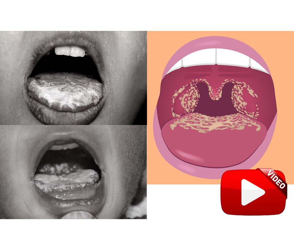
What is Oral Candidiasis? - Everything you need to know - Symptoms, diagnosis and treatment

Periodoncia

The Candida Albicans fungus, responsible for oral candidiasis, lives in our oral cavity together with other microorganisms but does not generate any pathological alteration.

📌 Recommended Article:
Download the PDF 🔽 Management of Angular Cheilitis in children ... Angular Cheilitis occurs more in children and it is caused by children sensitivity against certain contact agents like toys, foods, sunlight, allergy against medicines, cosmetics, and long term antibiotic treatment.

The disease manifests itself when the balance of the bacterial flora is disturbed or the patient's immunity decreases. Candida albicans most commonly affects infants and older adults, as they often have reduced immunity.

Advertisement

All the information about Oral Candidiasis and its causes, symptoms, diagnosis and treatment in the video that we share below.

📌 More recommended items

► Guide for the surgical management and oral pathology of the pediatric patient
► Deformity of the tongue in an infant: Riga-Fede disease
► How to Manage a Pediatric Patient with Oral Ulcers

📌 Watch the video "ORAL CANDIDIASIS | Diagnosis and Management "


Youtube/ TaughtWell SimplifyEd

lunes, 5 de octubre de 2020

Herpes labial y Candidiasis oral: ¿Cómo detectarlas?

Herpes

La cavidad bucal puede presentar una serie de afecciones, que pueden ser causadas por bacterias, virus u hongos. Entre las dos más comunes tenemos al herpes y a la candidiasis.

El herpes labial es producido por un virus (VHS-1), también existe un tipo de herpes genital producido por el VHS-2. La candidiasis es causada por un hongo (Candida Albicans), y se reconoce por manchas blancas en la mucosa oral.

Enlaces Patrocinados

Ambas afecciones se presentan de maneras diferentes, y es importante que se conozca el aspecto de cada una de ellas. La intervención profesional debe ser rápida y oportuna.

Medicina Bucal


También te puede interesar :
► ¿Es necesario extraer "las muelas del juicio" antes de un tratamiento de ortodoncia?
► Tratamiento de la mordida cruzada anterior con plano inclinado anterior. Efecto sobre los arcos dentales
► ¿Cómo se colocan los brackets?


Fuente: Youtube / El Heraldo de México
Imagen: GETTY//DIETMAR HÃÂPFL