Mostrando entradas con la etiqueta Article PDF. Mostrar todas las entradas

lunes, 6 de junio de 2022

Management of mandibular fractures in children. Diagnosis and treatment. Case report

Oral Surgery

Mandibular fractures in children are rare, and deserve special care because they are in full growth and craniofacial development. The appropriate treatment will depend on the age of the child and the affected mandibular area.

The evaluation and treatment must be immediate to avoid functional disorders, serious consequences in the craniofacial development and in the aesthetics of the patient. The treatment of mandibular fractures can be conservative or surgical.

Advertisement

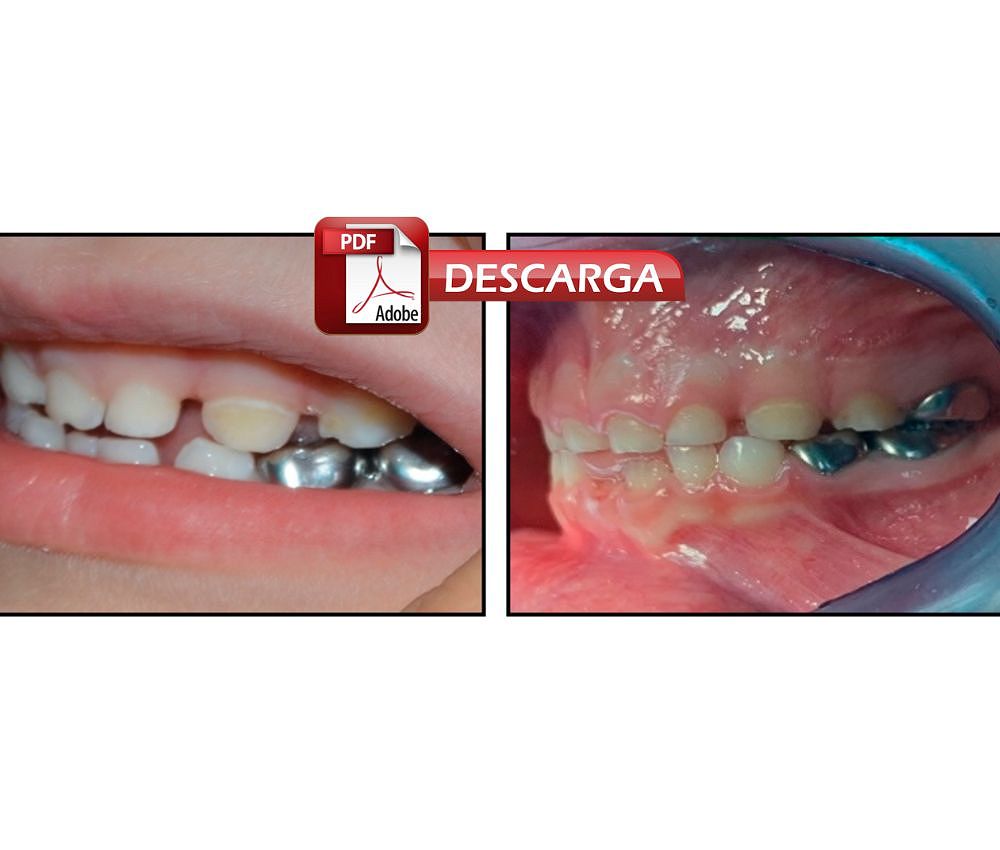
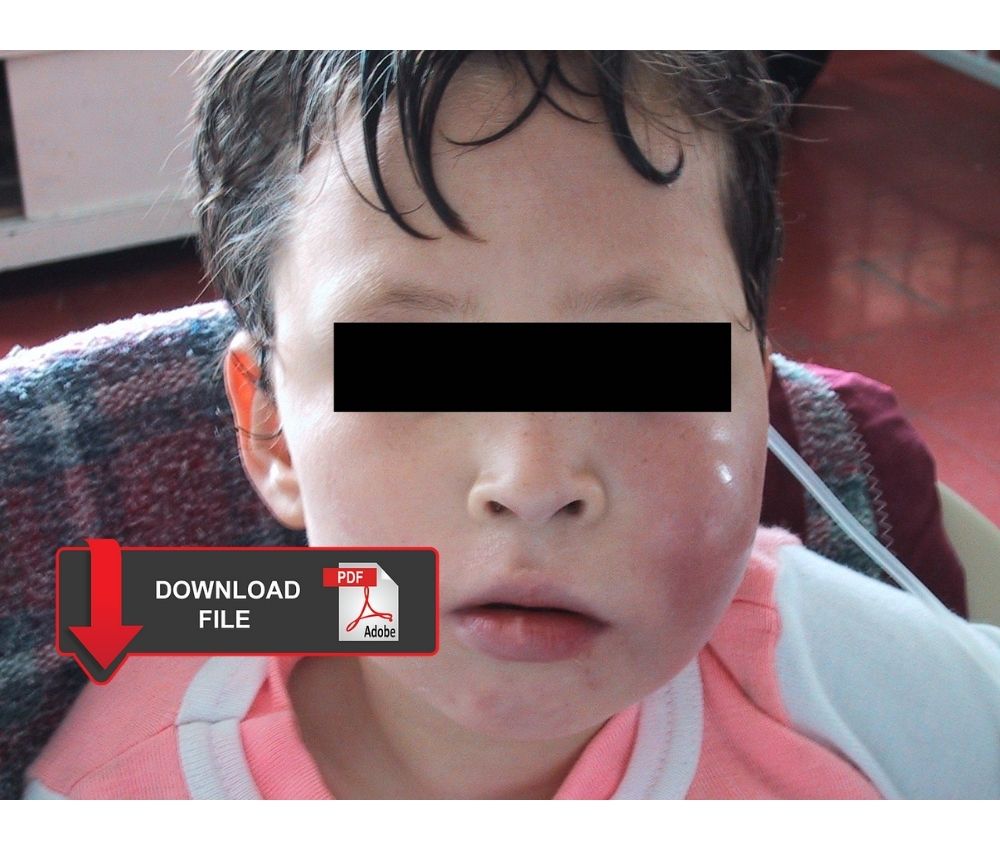
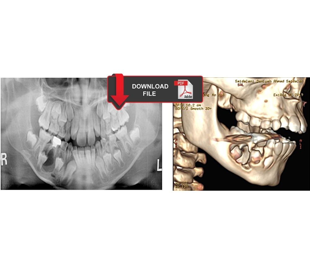
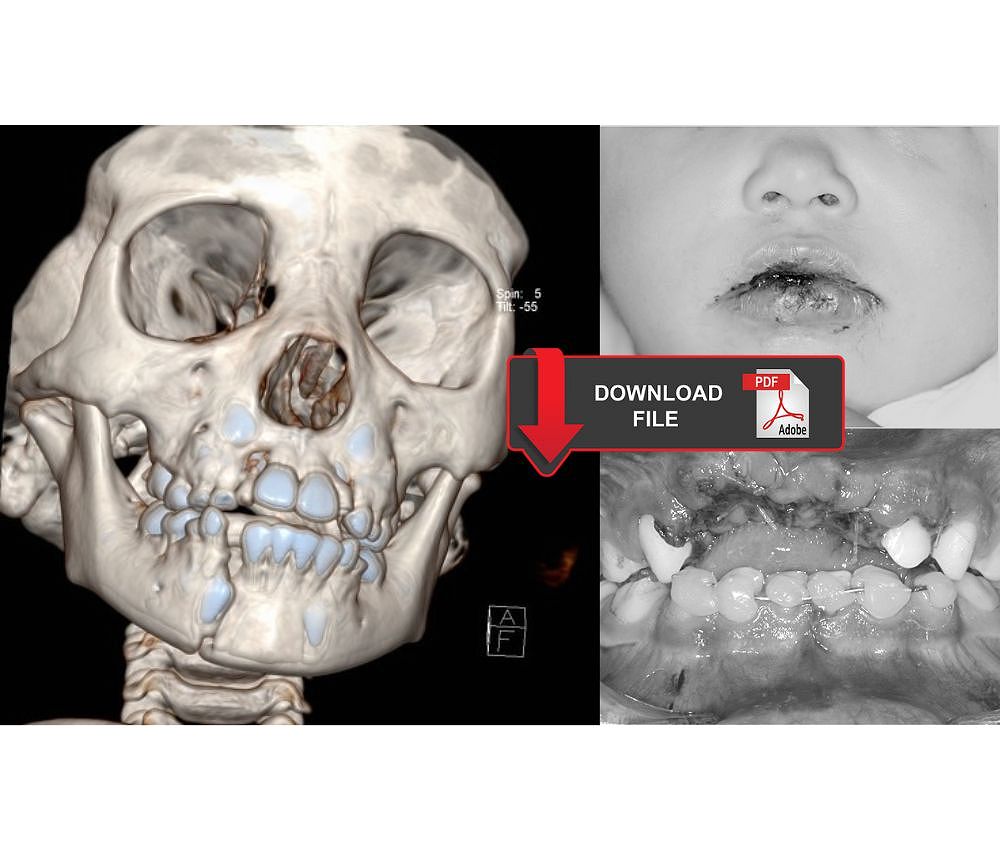
We share the recommendations for the diagnosis, management and treatment of mandibular fractures in pediatric patients, and we report the case of a 3-year-old patient and her surgical treatment.

Oral Surgery


👉 Article 1: READ AND DOWNLOAD "Mandibular fractures in children under 3 years: A rare case report" IN FULL IN PDF👈


Renato Maranoa, Patrício de Oliveira Netoa, Keiko Oliveira Sakugawab, Liliane S.S. Zanettic, Márcio de Moraesa. Mandibular fractures in children under 3 years: A rare case report. Rev Port Estomatol Med Dent Cir Maxilofac. 2013;54(3):166–170

👉 Article 2: READ AND DOWNLOAD "What are the differences in Pediatric Mandible Fractures?" IN FULL IN PDF👈


Karacor- Altuntas Z, Ismayilzade M (2017) What are the Differences in Pediatric Mandible Fractures? J. Aesthet Reconstr Surg. Vol 3 No.2: 11

You may also like :
Marsupialization of a large mandibular cyst in a pediatric patient - Clinical Case
Mandibular tumors in pediatric patients. Report of 04 cases of aggressive tumors
Reimplantation of avulsed permanent teeth after three days: Clinical case in a pediatric patient

viernes, 3 de junio de 2022

Natal teeth and Riga Fede ulcer: Diagnosis and treatment

Riga Fede

Riga Fede ulcer is usually located on the ventral side of the tongue, and is caused by the trauma caused by the presence of a natal or neonatal tooth. It is painful, so it limits the sucking action of breast milk, putting the baby's feeding at risk.

Natal and neonatal teeth are of unknown etiology, and are characterized by not presenting a single shape, they can be small or large and conical or normal in shape.

Advertisement

We share the clinical implications and the approach to the treatment of natal and neonatal teeth, regarding the case of the presence of Riga Fede ulcer in a two-month-old baby.

Riga-Fede Disease


👇 READ AND DOWNLOAD THE ARTICLE "Natal teeth and Riga Fede ulcer: Diagnosis and treatment" IN PDF 👇


Danelon, M., Emerenciano, N. G., Garcia, L. G., Percinoto, C., & Cunha, R. F. (2017). Natal teeth associated with Riga-Fede ulcer: case report. ARCHIVES OF HEALTH INVESTIGATION, 6(4). https://doi.org/10.21270/archi.v6i4.2059

You may also like :
Deformity of the tongue in an infant: Riga-Fede disease
Riga Fede Disease Treatment: Case Report
Guide for the surgical management and oral pathology of the pediatric patient

miércoles, 1 de junio de 2022

Hall technique: Complete information for the treatment of carious primary molars

Hall Technique

Caries is an infectious process that seriously affects dental enamel, and when it is not treated in time, it endangers the permanence of the tooth in the oral cavity.

Steel crowns are used in pediatric dentistry when primary teeth are severely damaged by dental caries. They are usually placed after pulp treatment and in hypoplastic teeth.

Advertisement

The objective of the article is to provide adequate information on the Hall technique, indications, contraindications, disadvantages, and the cost-effectiveness of this procedure. A complete analysis of this rehabilitation technique for carious primary teeth.

Hall Technique


👇 READ AND DOWNLOAD THE ARTICLE "Hall technique: Complete information for the treatment of carious primary molars" IN PDF 👇


Altoukhi DH, El-Housseiny AA. Hall Technique for Carious Primary Molars: A Review of the Literature. Dent J (Basel). 2020 Jan 17;8(1):11. doi: 10.3390/dj8010011. PMID: 31963463; PMCID: PMC7148518.

You may also like :
The Hall Technique: Manual for the management of primary molar caries
Preparation of stainless steel crown for the rehabilitation of primary molars
Preformed crowns for decayed primary molar teeth (Review)

martes, 31 de mayo de 2022

Dentigerous cyst in mixed and primary dentition. Diagnosis and surgical treatment

Dentigerous cysts

Dentigerous cysts are cavities with liquid content, and develop from the remains of the enamel epithelium. They are the most common, benign cysts, and are usually located in the lower jaw. It is related to the crowns of unerupted teeth.

They manifest as radiographic findings or when the patient has acute inflammation. Enucleation of the cyst is performed using the surgical technique of marsupialization.

Advertisement

We share several cases that report the diagnosis, management and surgical treatment (enucleation and marsupialization) of the dentigerous cyst in mixed and primary dentition.

Oral Surgery


👉 ARTICLE 1: READ AND DOWNLOAD "Dentigerous Cysts in Primary Dentition: Report of 2 Cases" IN FULL IN PDF👈


Delbem AC, Cunha RF, Afonso RL, Bianco KG, Idem AP. Dentigerous cysts in primary dentition: report of 2 cases. Pediatr Dent. 2006 May-Jun;28(3):269-72. PMID: 16805361.

👉 ARTICLE 2: READ AND DOWNLOAD "Dentigerous cyst in a young child: a case report" IN FULL IN PDF👈


McKinney, S. L., & Lukes, S. M. (2021). Dentigerous cyst in a young child: a case report. Canadian journal of dental hygiene : CJDH = Journal canadien de l'hygiene dentaire : JCHD, 55(3), 177–181.

👉 ARTICLE 3: READ AND DOWNLOAD "Dentigerous Cyst in Children: A Case Report and Outline of Clinical Management for Pediatric and General Dentists" IN FULL IN PDF👈


Meer, Zakirulla & Yavagal, Chandrashekar & Jayashankar, DN & Meer, Allahbaksh. (2012). Dentigerous Cyst in Children: A Case Report and Outline of Clinical Management for Pediatric and General Dentists. Journal of Orofacial Research. 2. 238-242. 10.5005/jp-journals-10026-1051.

You may also like :
Guide for the surgical management and oral pathology of the pediatric patient
Marsupialization of a large mandibular cyst in a pediatric patient - Clinical Case
Management of acute orofacial infection of odontogenic origin in children - Diagnosis, clinic and pharmacology

martes, 24 de mayo de 2022

Marsupialization of a large mandibular cyst in a pediatric patient - Clinical Case

Oral Surgery

Mandibular cysts are generally benign neoplasms, may be of odontogenic origin and vary in size and severity. Mandibular cysts can expand, injuring large extensions of bone tissue, involving the stability of the teeth.

Various techniques are used to surgically remove the mandibular cyst, enucleation when the cyst is small, or marsupialization when it is large.

Advertisement

We share the case of surgical removal (marsupialization) of a large mandibular cyst in a pediatric patient, and the use of a multipurpose space maintainer.

Oral Surgery


👉 READ AND DOWNLOAD "Marsupialization of a large mandibular cyst in a pediatric patient - Clinical Case" IN FULL IN PDF👈


Salem Waly. Department of Oral and Maxillofacial Surgery, Faculty of Dental Medicine, Al-Azhar University, Cairo, Egypt. 2090-097X, October 2019, Vol. 10, No. 4

You may also like :
Pharmacological treatment of oral infections in pediatric dentistry - Recommendations and dose calculation
Mandibular tumors in pediatric patients. Report of 04 cases of aggressive tumors
Management of acute orofacial infection of odontogenic origin in children - Diagnosis, clinic and pharmacology

jueves, 19 de mayo de 2022

What are those bony bumps in my mouth? (Torus)

Torus

Torus are exostoses or benign bony protrusions that appear in the upper or lower jaw. It can be unilateral or bilateral, and its etiology is unknown, but it is related to genetic factors, trauma or environmental factors.

The torus should be removed if they are large and generate aesthetic, chewing, swallowing problems, or when they interfere with the making of dental prostheses.

Advertisement

The bony bumps (torus) are slow growing and can appear in the upper or lower jaw, their removal is surgical. More information about its characteristics and manifestations in the article that we share.

Oral Surgery


👉 READ AND DOWNLOAD "What are those bony bumps in my mouth?" IN FULL IN PDF👈


Soumya Chatterjee. Cleveland Clinic Journal of Medicine Jan 2016, 83 (1) 17-18; DOI: 10.3949/ccjm.83a.15033

You may also like :
What is pre-prosthetic surgery?
Tooth Extraction: Post-Surgical Care and Tips
How is a palatine torus removed? - Step by Step

lunes, 16 de mayo de 2022

Oral manifestations of eating disorders in adolescent patients

Oral Medicine

Eating disorders are a set of psychopathological disorders that are related to eating behaviors, and affect physical and emotional health, these conditions usually manifest in young people and adolescents. The most common eating disorders are anorexia and bulimia.

Patients with eating disorders present alterations in the perioral tissues, oral mucosa and teeth. These alterations generate several problems in the different functions of the oral cavity.

Advertisement

We share a study that describes the different oral manifestations of eating disorders (anorexia, bulimia) that we can find in an adolescent patient with the aim that the professional identifies these signs early.

Oral Medicine


👉 READ AND DOWNLOAD "Odontogenic facial cellulitis in a pediatric patient - Medical-dental management considerations" IN FULL IN PDF👈


Monda M, Costacurta M, Maffei L, Docimo R. Oral manifestations of eating disorders in adolescent patients. A review. Eur J Paediatr Dent. 2021 Jun;22(2):155-158. doi: 10.23804/ejpd.2021.22.02.13

You may also like :
How to identify the early signs of oral cancer
Bruxism: A medical or dental issue?
Allergic manifestations to local anaesthetic in pediatric dentistry: Prevention and management