Mostrando entradas con la etiqueta Fluorosis. Mostrar todas las entradas

sábado, 21 de febrero de 2026

Stained Teeth in Children: Common Causes and Safe Esthetic Treatments

Stained Teeth

Dental discoloration in children is a frequent concern for parents and clinicians, often affecting both esthetics and psychosocial well-being.

📌 Recommended Article :
Dental Article 🔽 Black Stains on Primary Teeth in Children: Causes, Diagnosis, and Management ... Understanding their etiology, clinical presentation, and management is essential to avoid overtreatment and to provide accurate preventive guidance.
Pediatric tooth staining may result from developmental enamel defects, systemic conditions, or environmental factors, and requires an accurate diagnosis to ensure safe and effective management.

Advertisement

This article reviews the most common causes of stained teeth in children, including fluorosis, enamel hypoplasia, and other pathologies, and discusses modern, minimally invasive esthetic treatments, such as remineralization therapies and microabrasion, based on current scientific evidence.

Common Causes of Stained Teeth in Children

1. Dental Fluorosis
Dental fluorosis is a developmental condition caused by excessive fluoride intake during enamel formation. Clinically, it presents as white opacities, yellow-brown stains, or surface porosities, depending on severity.

▪️ Typically symmetrical
▪️ Affects permanent teeth
▪️ Esthetic impact varies from mild to severe

2. Enamel Hypoplasia and Hypomineralization
Enamel hypoplasia results from quantitative defects in enamel formation, while hypomineralization reflects qualitative changes.
Common features include:

▪️ Demarcated white, yellow, or brown defects
▪️ Increased caries susceptibility
▪️ Rough or pitted enamel surfaces
Conditions such as molar-incisor hypomineralization (MIH) fall within this category and are increasingly reported worldwide.

3. Other Pathologies and Extrinsic Factors
Additional causes of discoloration include:

▪️ Early childhood caries (ECC) leading to dark or chalky lesions
▪️ Chromogenic bacteria, associated with black line stains
▪️ Trauma to primary teeth, causing intrinsic discoloration of successors
▪️ Medications, such as tetracyclines (rare but relevant in historical cases)
▪️ Accurate differentiation is essential to avoid overtreatment.

📌 Recommended Article :
Dental Article 🔽 Enamel Hypoplasia vs Dental Fluorosis: Key Differences, Diagnosis, and Treatment ... While both conditions alter enamel structure and appearance, their etiology, presentation, and management differ significantly.
Modern Esthetic and Conservative Treatment Options

1. Remineralization Therapies
Contemporary pediatric dentistry prioritizes non-invasive remineralization strategies, especially for early enamel defects.

These include:
▪️ Fluoride varnishes
▪️ Casein phosphopeptide–amorphous calcium phosphate (CPP-ACP)
▪️ Calcium phosphate-based agents
▪️ Silver diamine fluoride (selected cases)
These approaches are safe, effective, and suitable for young patients.

2. Enamel Microabrasion
Microabrasion is indicated for superficial intrinsic stains, particularly mild fluorosis.
Advantages include:

▪️ Conservative enamel removal
▪️ Immediate esthetic improvement
▪️ Long-term stability when correctly indicated
It is often combined with remineralization to enhance outcomes.

3. Resin Infiltration and Restorative Options
For deeper lesions, resin infiltration or minimally invasive restorations may be considered, always balancing esthetics with tooth preservation.

📌 Recommended Article :
Dental Article 🔽 Molar-Incisor Hypomineralization and Enamel Hypoplasia: Updated Clinical Approaches in Pediatric Dentistry ... Differentiating them is critical, as each condition requires a distinct diagnostic and therapeutic approach. This article presents the latest scientific evidence on their definition, etiology, diagnosis, and modern management.
💬 Discussion
The management of stained teeth in children requires a precise differential diagnosis, as treatment effectiveness depends on the underlying etiology. Advances in preventive and minimally invasive dentistry allow clinicians to address esthetic concerns while preserving tooth structure and ensuring safety.
Misdiagnosis may lead to inappropriate interventions, highlighting the importance of clinical expertise and evidence-based decision-making.

🎯 Clinical Recommendations
▪️ Perform thorough clinical and radiographic assessment
▪️ Identify whether stains are developmental, acquired, intrinsic, or extrinsic
▪️ Prioritize remineralization and minimally invasive techniques
▪️ Educate parents regarding etiology and realistic outcomes
▪️ Avoid aggressive esthetic treatments in young patients

✍️ Conclusion
Stained teeth in children are multifactorial and require individualized management. Conditions such as fluorosis and enamel hypoplasia can be effectively managed using safe, conservative, and modern esthetic approaches, including remineralization and microabrasion. Early diagnosis and appropriate treatment planning are essential to achieve optimal functional and esthetic outcomes.

📊 Comparative Table: Differential Diagnosis of Pediatric Tooth Staining

Condition Key Clinical Features Diagnostic Limitations
Dental Fluorosis Symmetrical white or brown opacities Severity may be underestimated clinically
Enamel Hypoplasia Localized pits, grooves, or missing enamel May resemble post-eruptive breakdown
MIH Demarcated opacities on molars and incisors Variable severity complicates diagnosis
Extrinsic Staining Black or brown surface stains Easily confused with early caries
📚 References

✔ American Academy of Pediatric Dentistry. (2023). Guideline on management of dental patients with enamel defects. Pediatric Dentistry, 45(6), 315–322.
✔ Fejerskov, O., Nyvad, B., & Kidd, E. (2015). Dental caries: The disease and its clinical management (3rd ed.). Wiley Blackwell.
✔ Gugnani, N., Pandit, I. K., Gupta, M., & Gugnani, S. (2017). Esthetic management of fluorosis in children. Journal of Esthetic and Restorative Dentistry, 29(5), 303–312. https://doi.org/10.1111/jerd.12312
✔ Weerheijm, K. L. (2018). Molar-incisor hypomineralisation (MIH). European Archives of Paediatric Dentistry, 19(4), 225–232. https://doi.org/10.1007/s40368-018-0354-9

📌 More Recommended Items

Enamel Erosion: Causes, Symptoms, Prevention, and Treatment for a Healthy Smile
Fluoride Varnish for Preventing and Treating White Spot Lesions: Clinical Evidence and Best Practices
SMART Technique in Pediatric Dentistry: Caries Management in Children

domingo, 15 de febrero de 2026

Fluorosis vs. Enamel Demineralization: How to Identify Clinical Differences and Choose the Right Treatment

Fluorosis - Enamel Demineralization

Dental fluorosis and enamel demineralization are common enamel alterations that may present with similar clinical appearances but differ significantly in etiology, progression, and management.

📌 Recommended Article :
Dental Article 🔽 Fluoride in Pediatric Dentistry: Dosage, Frequency, and Current Controversies ... Managing the behavior of the pediatric patient is a challenge for the pediatric dentist, since attending the office generates stress and uncertainty, causing some type of behavioral response.
Accurate differentiation is essential to avoid misdiagnosis and to implement appropriate preventive or therapeutic strategies. This article reviews the clinical characteristics, diagnostic considerations, and current treatment approaches for fluorosis and demineralization, providing practical guidance for dental professionals.

Advertisement

Introduction
White enamel defects are frequently observed in both pediatric and adult dental patients. Among these, fluorosis and enamel demineralization represent two distinct pathological processes. While fluorosis is a developmental condition linked to excessive fluoride intake during enamel formation, demineralization is an acquired process related to biofilm activity and acid exposure. Misinterpretation may lead to ineffective or unnecessary treatment, highlighting the importance of proper clinical differentiation.

📌 Recommended Article :
Dental Article 🔽 Enamel Hypoplasia vs Molar-Incisor Hypomineralization (MIH): Diagnosis and Modern Management ... This article explores their etiology, clinical characteristics, and modern treatment options, providing a comprehensive guide for accurate diagnosis and management.
1. Dental Fluorosis

Etiology
Dental fluorosis results from excessive systemic fluoride ingestion during amelogenesis, typically before the age of eight. The excess fluoride disrupts enamel mineralization, leading to increased porosity.

Clinical Characteristics
▪️ Symmetrical distribution affecting homologous teeth
▪️ Diffuse white opacities, striations, or mottling
▪️ In moderate to severe cases, brown discolorations and surface pitting
▪️ Lesions are present at eruption and do not progress over time

Treatment Options
Management depends on severity:
▪️ Mild fluorosis: No treatment or enamel polishing
▪️ Moderate fluorosis: Resin infiltration or microabrasion
▪️ Severe fluorosis: Composite restorations or porcelain veneers

📌 Recommended Article :
Dental Article 🔽 Minimally Invasive Pediatric Dentistry: Hall Technique, SMART, and ART — A Clinical Guide for Parents and Practitioners ... Modern approaches such as the Hall Technique, SMART, and ART offer evidence-based alternatives that avoid drilling, reduce fear, and improve cooperation in young patients.
2. Enamel Demineralization

Etiology
Enamel demineralization is caused by prolonged exposure to acids produced by cariogenic bacteria. It commonly occurs around orthodontic brackets or in areas of poor oral hygiene.

Clinical Characteristics
▪️ Chalky white spot lesions with well-defined borders
▪️ Typically localized and asymmetrical
▪️ Lesions develop after tooth eruption
▪️ May progress to cavitated carious lesions if untreated

Treatment Options
▪️ Topical fluoride therapy (varnishes, gels)
▪️ Casein phosphopeptide–amorphous calcium phosphate (CPP-ACP)
▪️ Resin infiltration for non-cavitated lesions
▪️ Restorative treatment if cavitation is present

📊 Comparative Table: Fluorosis vs. Enamel Demineralization

Clinical Parameter Fluorosis Enamel Demineralization
Time of Onset During tooth development After tooth eruption
Lesion Distribution Symmetrical and generalized Localized and asymmetrical
Progression Non-progressive Progressive if untreated
Primary Management Esthetic-focused treatment Remineralization and caries control
💬 Discussion
Although both conditions present as white enamel defects, fluorosis is a developmental and non-progressive condition, whereas demineralization is an active, biofilm-mediated process. The timing of lesion appearance, symmetry, and surface texture are critical diagnostic clues. Adjunctive tools such as quantitative light-induced fluorescence (QLF) may support diagnosis but do not replace thorough clinical examination.

🎯 Clinical Recommendations
▪️ Always assess patient age and fluoride exposure history
▪️ Evaluate lesion symmetry, borders, and location
▪️ Avoid aggressive treatment in mild fluorosis
▪️ Implement early remineralization protocols for demineralization
▪️ Educate patients and caregivers on fluoride use and oral hygiene

✍️ Conclusion
Distinguishing fluorosis from enamel demineralization is essential for accurate diagnosis and appropriate management. Understanding their distinct etiologies and clinical behaviors allows clinicians to deliver targeted, conservative, and effective treatments, improving both functional and esthetic outcomes.

📚 References

✔ Fejerskov, O., Nyvad, B., & Kidd, E. (2015). Dental caries: The disease and its clinical management (3rd ed.). Wiley-Blackwell.
✔ Ismail, A. I., Sohn, W., Tellez, M., Amaya, A., Sen, A., Hasson, H., & Pitts, N. B. (2013). The International Caries Detection and Assessment System (ICDAS). Community Dentistry and Oral Epidemiology, 41(1), e41–e53. https://doi.org/10.1111/cdoe.12006
✔ Pendrys, D. G. (2010). Risk of enamel fluorosis in nonfluoridated and fluoridated communities. Journal of Public Health Dentistry, 70(2), 79–85. https://doi.org/10.1111/j.1752-7325.2010.00164.x
✔ Pretty, I. A. (2006). Caries detection and diagnosis. Dental Clinics of North America, 50(4), 515–527. https://doi.org/10.1016/j.cden.2006.06.003

📌 More Recommended Items

Fluoride Toxicity in Children: Symptoms, Immediate Actions, and Prevention — A Comprehensive Clinical Guide
What Is MIH? Understanding Weak Enamel in Children’s Teeth and How to Treat It
Understanding Dental Erosion: Causes and Prevention (Updated Guide)

miércoles, 3 de diciembre de 2025

Fluoride Toxicity in Children: Symptoms, Immediate Actions, and Prevention — A Comprehensive Clinical Guide

Fluoride Toxicity

Fluoride toxicity in children remains a significant clinical concern due to the widespread use of fluoride-containing products in pediatric oral health. While fluoride is essential for dental caries prevention, excessive ingestion can lead to acute or chronic toxicity, compromising systemic health.

📌 Recommended Article :
Dental Article 🔽 Fluoride in Pediatric Dentistry: Dosage, Frequency, and Current Controversies ... Key aspects include topical vs. systemic fluoride, caries-preventive effectiveness, and a special update on recent FDA considerations (2025) related to systemic fluoride supplements.
This article provides an updated, evidence-based, and SEO-optimized review of symptoms, emergency actions, preventive strategies, and comprehensive management of fluoride toxicity in the pediatric population.

Advertisement

Overview of Fluoride Toxicity
Fluoride toxicity occurs when the ingested dose exceeds the body’s capacity to metabolize and excrete fluoride safely. The probable toxic dose (PTD) is approximately 5 mg/kg body weight, and ingestion above this threshold requires emergency assessment (AAPD, 2023).
Excessive exposure can arise from toothpaste, mouth rinses, dietary supplements, or accidental ingestion of professional topical fluoride products.

📌 Recommended Article :
Dental Article 🔽 Silver Diamine Fluoride in Pediatric Dentistry: A Review ... Silver Diamine Fluoride (SDF) has emerged as a non-invasive and cost-effective treatment for managing dental caries in children.
How Fluoride Toxicity Affects General Health

➤ Systemic Impact
Excessive fluoride affects multiple body systems:
▪️ Gastrointestinal system: irritation of gastric mucosa causing nausea, vomiting, abdominal pain.
▪️ Electrolyte balance: risk of hypocalcemia and hyperkalemia due to fluoride’s strong affinity for calcium ions.
▪️ Neuromuscular system: muscle spasms, paresthesia, and in severe cases, seizures.
▪️ Cardiovascular system: altered cardiac function from electrolyte imbalance.
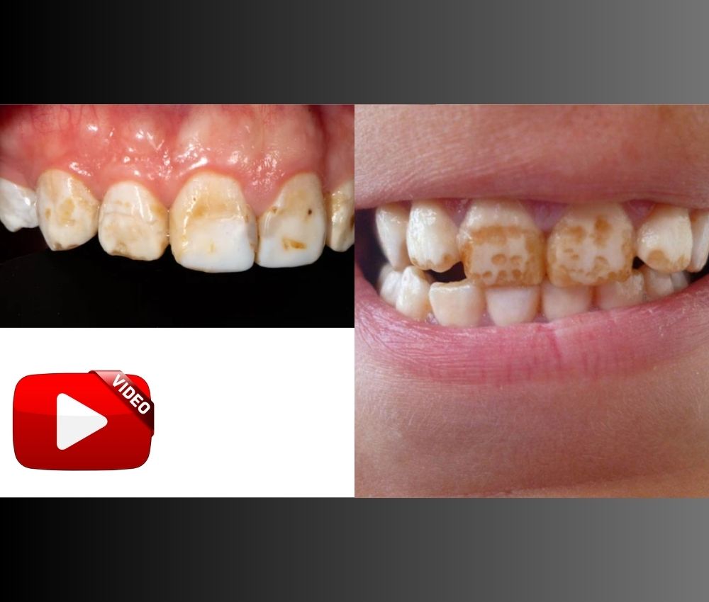
▪️ Skeletal development: chronic high intake may result in dental fluorosis and skeletal fluorosis.

📌 Recommended Article :
Dental Article 🔽 Fluoride Varnish in Pediatric Dentistry: Benefits, Indications, Mechanism, and Application Protocol ... Due to its ease of application, safety, and efficacy, it has become an essential part of caries management in clinical pediatric practice.
Early Signs and Symptoms of Fluoride Toxicity in Children

➤ Mild to moderate ingestion may present with:
▪️ Nausea and vomiting (often the earliest sign)
▪️ Hypersalivation
▪️ Abdominal pain
▪️ Diarrhea
▪️ Headache
▪️ Weakness or fatigue

➤ Severe ingestion may lead to:
▪️ Hypocalcemia-related tetany
▪️ Cardiac arrhythmias
▪️ Seizures
▪️ Respiratory depression
▪️ Life-threatening toxicity if untreated
Children are at higher risk because of lower body weight and tendency to swallow toothpaste.

📌 Recommended Article :
Dental Article 🔽 Enamel Hypoplasia vs Dental Fluorosis: Key Differences, Diagnosis, and Treatment ... Enamel hypoplasia and dental fluorosis are two developmental defects of enamel frequently encountered in clinical dentistry.
Immediate Actions When Fluoride Toxicity Is Suspected

➤ Emergency Management Protocol
1. Assess the amount ingested and body weight to estimate toxicity risk.
2. Administer milk or calcium-rich products immediately to bind fluoride and reduce absorption.
3. Do NOT induce vomiting.
4. Seek emergency medical evaluation, especially if ingestion approaches or exceeds 5 mg/kg.
5. Monitor vital signs and prepare for electrolyte correction in a clinical setting.
6. Severe cases may require IV calcium gluconate, cardiac monitoring, and supportive therapy.

📌 Recommended Article :
Webinar 🔽 Webinar: Silver Diamine Fluoride – A Silver Renaissance in Dentistry - Dr. Anas Al Salami ... Silver Diamine Fluoride (SDF) has emerged as a transformative option in pediatric dentistry, marking a true renaissance in caries management.
Prevention Strategies in Pediatric Dental Care

➤ Evidence-Based Prevention Measures
▪️ Parents should supervise brushing until at least age 6.
▪️ Use smear layer of toothpaste for children under 3; use a pea-sized amount from ages 3–6 (CDC, 2024).
▪️ Store fluoride products out of children’s reach.
▪️ Avoid flavored toothpaste that encourages swallowing.
▪️ Evaluate dietary fluoride sources: water fluoridation, supplements, combined exposures.
▪️ Provide structured parental counseling during dental visits.

📊 Comparative Table: Fluoride Sources in Children

Aspect Advantages Limitations
Community Water Fluoridation Reduces caries at population level Risk of overexposure if combined with supplements
Fluoride Toothpaste Highly effective; easy to access Children may swallow excessive amounts if unsupervised

💬 Discussion
The balance between the preventive benefits of fluoride and the risks of toxicity requires strategic dosing, parental education, and clinician guidance. Most toxicity events are preventable and relate to unsupervised access or incorrect use of fluoride-containing products. Updated protocols from ADA and CDC emphasize minimizing risk through tailored dosing and behavioral supervision.

📌 Recommended Article :
PDF 🔽 Fluoride Varnish in the Prevention of Dental Caries in Children and Adolescents: A Systematic Review ... We share an article that reviews the literature and develops a protocol for the use of fluoride varnish as a caries preventive agent in children and adolescents.
✍️ Conclusion
Fluoride toxicity in children is rare but potentially serious. Early recognition of symptoms, prompt emergency actions, and preventive strategies are crucial to ensuring safety. When used correctly, fluoride remains one of the most powerful tools for caries prevention in pediatric dentistry. Clinicians must continue reinforcing safe use to eliminate avoidable toxic exposures.

🔎 Recommendations
▪️ Educate parents on appropriate fluoride dosing based on age.
▪️ Emphasize supervision during brushing.
▪️ Assess total fluoride exposure at every dental visit.
▪️ Maintain updated emergency protocols for accidental ingestion.
▪️ Promote community awareness regarding the safe storage of dental products.

📚 References

✔ American Academy of Pediatric Dentistry. (2023). Policy on use of fluoride. AAPD Reference Manual. https://www.aapd.org
✔ Centers for Disease Control and Prevention. (2024). Children’s oral health: Fluoride use. U.S. Department of Health and Human Services. https://www.cdc.gov
✔ Agency for Toxic Substances and Disease Registry. (2023). Toxicological profile for fluoride. U.S. Department of Health and Human Services. https://www.atsdr.cdc.gov
✔ Whitford, G. M. (2011). Fluoride in dental products: Safety considerations. Journal of Dental Research, 90(6), 573–582. https://doi.org/10.1177/0022034510384626

📌 More Recommended Items

Latest FDA Guidelines (2025) on Fluoride Use in Children: What Dentists Need to Know
Xylitol and Dental Caries Prevention in Children: Mechanism, Benefits, and Clinical Use
Fluoride Recommendations for Children in the US: What Parents Should Know

miércoles, 22 de octubre de 2025

White or Brown Spots on Teeth? Understanding Fluorosis and Enamel Hypoplasia

Fluorosis - Enamel Hypoplasia

Introduction
White or brown spots on teeth are among the most common esthetic concerns in both children and adults. Two main conditions often responsible for these enamel defects are dental fluorosis and enamel hypoplasia.

📌 Recommended Article :
Dental Article 🔽 Enamel Hypoplasia vs Dental Fluorosis: Key Differences, Diagnosis, and Treatment ... While both conditions alter enamel structure and appearance, their etiology, presentation, and management differ significantly. Understanding these differences is essential for accurate diagnosis and effective treatment planning.
Although they may appear similar, their etiology, diagnosis, and management differ significantly. Correct differentiation is essential for successful treatment and patient satisfaction.

Advertisement


Definition and Etiology

➤ Dental Fluorosis
Dental fluorosis is a developmental disturbance of enamel caused by excessive fluoride ingestion during tooth formation (typically before age 8). Fluoride interferes with ameloblast activity, leading to hypomineralized enamel.
▪️ Mild fluorosis manifests as faint white lines or cloudy opacities.
▪️ Moderate to severe fluorosis presents as brown discoloration, surface irregularities, and in extreme cases, enamel pitting.
| Common sources include fluoridated water, toothpaste ingestion, and fluoride supplements.

➤ Enamel Hypoplasia
Enamel hypoplasia is a quantitative defect of enamel formation, resulting from disruption in ameloblast function during enamel matrix secretion. It leads to thin or missing enamel areas, with visible grooves, pits, or chalky opacities.
Etiologic factors include:
▪️ Nutritional deficiencies (Vitamin D, calcium)
▪️ Infections (measles, chickenpox) during tooth formation
▪️ Premature birth or low birth weight
▪️ Trauma or systemic diseases affecting amelogenesis

📌 Recommended Article :
Dental Article 🔽 Enamel Hypoplasia vs Molar-Incisor Hypomineralization (MIH): Diagnosis and Modern Management ... Enamel defects are among the most common developmental disturbances in pediatric dentistry. Two major entities—enamel hypoplasia and molar-incisor hypomineralization (MIH)—are often confused due to overlapping clinical features.
Differential Diagnosis
Distinguishing between fluorosis and hypoplasia is essential.

▪️ Fluorosis: Symmetrical, diffuse opacities without enamel loss.
▪️ Hypoplasia: Asymmetrical, well-defined defects with enamel reduction.
Diagnostic tools include:
▪️ Clinical examination using transillumination and drying techniques.
▪️ Patient history regarding fluoride exposure or childhood illnesses.
▪️ Photographic documentation and DIAGNOdent laser fluorescence can aid in differential identification

📌 Recommended Article :
PDF/Video 🔽 Molar-Incisor Hypomineralization: Clinical Manifestations and Treatment ... Clinically, Molar Incisor Hypomineralization is characterized by presenting a dental piece with an enamel that fractures easily and of various colors that vary between yellow, brown and white.
Modern Treatment Options
Treatment depends on the severity, esthetic concern, and patient age.
Conservative treatments include:
▪️ Microabrasion to remove superficial stains.
▪️ Resin infiltration (ICON®) to mask white lesions and harmonize color.
▪️ Topical remineralization with CPP-ACP (casein phosphopeptide–amorphous calcium phosphate) or fluoride varnish to improve surface hardness.
Advanced esthetic treatments for moderate to severe cases:
▪️ Composite resin restorations for small defects.
▪️ Porcelain veneers or full crowns for extensive enamel loss.
▪️ Bleaching protocols may be used carefully in mild fluorosis to improve color uniformity.
Modern digital dentistry tools, such as AI-based color mapping and minimally invasive laser techniques, are enhancing accuracy and esthetic outcomes.

📊 Comparative Table: Modern Treatments for Fluorosis and Enamel Hypoplasia

Aspect Advantages Limitations
Microabrasion Minimally invasive; improves mild discoloration effectively Limited depth removal; not effective for deep defects
Resin Infiltration (ICON®) Camouflages white spots; preserves healthy enamel Costly; requires high operator skill
Topical Remineralization (CPP-ACP, Fluoride) Non-invasive; strengthens enamel and prevents progression Results are gradual; limited esthetic improvement
Composite Restorations Immediate esthetic correction; customizable shade May discolor or wear over time; technique sensitive
Porcelain Veneers/Crowns Excellent esthetics; durable long-term outcome Invasive; higher cost and irreversible

✍️ Conclusion
Fluorosis and enamel hypoplasia share similar visual characteristics but differ in origin and clinical implications. Accurate diagnosis allows clinicians to select conservative, evidence-based treatments that maintain tooth structure while improving esthetics. The integration of minimally invasive techniques, digital tools, and remineralization therapies provides predictable, patient-centered outcomes.

📌 Recommended Article :
Dental Article 🔽 Enamel Erosion: Causes, Symptoms, Prevention, and Treatment for a Healthy Smile ... Enamel erosion is a dental condition characterized by the progressive loss of the tooth's outermost layer, known as enamel. This irreversible process can lead to various oral health issues if not addressed promptly.
🔎 Recommendations
▪️ Perform a detailed fluoride exposure history for every pediatric patient.
▪️ Use non-invasive treatments first, reserving restorations for severe cases.
▪️ Educate caregivers about optimal fluoride use and nutrition during tooth development.
▪️ Schedule periodic follow-ups to monitor enamel stability and esthetic satisfaction.

📚 References

✔ Aoba, T., & Fejerskov, O. (2002). Dental fluorosis: chemistry and biology. Critical Reviews in Oral Biology & Medicine, 13(2), 155–170. https://doi.org/10.1177/154411130201300206
✔ Crombie, F. A., Manton, D. J., & Palamara, J. E. (2013). Comparison of the mechanical properties of hypomineralised enamel and normal enamel. Journal of Dentistry, 41(2), 135–142. https://doi.org/10.1016/j.jdent.2012.11.002
✔ El Mourad, A. M. (2018). Aesthetic management of enamel hypoplasia and fluorosis: conservative approaches. Journal of Clinical and Experimental Dentistry, 10(9), e896–e903. https://doi.org/10.4317/jced.54920
✔ Wong, H. M., & McGrath, C. (2014). Esthetic perception and psychosocial impact of enamel defects among young adults. American Journal of Orthodontics and Dentofacial Orthopedics, 145(2), 191–199. https://doi.org/10.1016/j.ajodo.2013.10.015

📌 More Recommended Items

SMART Technique in Pediatric Dentistry: Caries Management in Children
Use of Silver Diamine Fluoride (SDF) - General Guide on its application
Glass Ionomer Cement in Pediatric Dentistry: Benefits, Uses, and Step-by-Step Application Guide

lunes, 20 de octubre de 2025

How to Identify and Treat Enamel Hypoplasia and Fluorosis in Dental Practice

Enamel Hypoplasia and Fluorosis

Abstract
Enamel hypoplasia and dental fluorosis are two prevalent developmental enamel defects that challenge both diagnosis and esthetic management in clinical dentistry.

📌 Recommended Article :
Dental Article 🔽 Chemical Caries Removal: Drill-Free Technique, Materials, and Clinical Application ... Chemical caries removal is a minimally invasive technique that allows for the selective elimination of decayed dentin without the use of a dental drill.
This article provides an evidence-based review of their etiology, clinical features, differential diagnosis, and modern treatment modalities, helping clinicians improve accuracy and patient outcomes.

Advertisement


Introduction
Developmental defects of enamel (DDE) are among the most frequent conditions affecting tooth structure in children. Enamel hypoplasia results from a quantitative defect in enamel formation, whereas fluorosis is a qualitative alteration caused by excessive fluoride intake during amelogenesis. Differentiating between these two is essential for accurate diagnosis, preventive counseling, and esthetic restoration.

Definition

➤ Enamel Hypoplasia: A quantitative defect in which the enamel thickness is reduced due to a disturbance during the secretory phase of amelogenesis (Suckling, 1989). Clinically, it appears as pits, grooves, or missing enamel.
➤ Dental Fluorosis: A qualitative defect resulting from excessive fluoride exposure during enamel maturation, leading to subsurface porosity and opacity (DenBesten & Li, 2011).

Etiology

➤ Enamel Hypoplasia
▪️ Prenatal causes: Maternal malnutrition, infections (rubella, syphilis), or systemic illness.
▪️ Perinatal causes: Birth trauma, hypoxia, or prematurity.
▪️ Postnatal causes: Fever, systemic diseases (measles, rickets), malnutrition, or trauma to primary teeth affecting successors.

➤ Dental Fluorosis
▪️ Chronic fluoride ingestion above 0.05 mg/kg/day during tooth development.
▪️ Sources include: Drinking water, toothpaste ingestion, and dietary supplements.
▪️ Severity correlates with fluoride concentration, exposure duration, and age.

Diagnosis

➤ Clinical Examination
Enamel hypoplasia manifests as well-demarcated pits, grooves, or missing enamel, while fluorosis appears as diffuse white, yellow, or brown opacities with symmetrical distribution.

➤ Radiographic Findings
▪️ Hypoplasia: Reduced enamel thickness and irregular surface.
▪️ Fluorosis: Normal enamel thickness but altered translucency.

✅ Differential Diagnosis Table

📊 Comparative Table: Enamel Hypoplasia vs Dental Fluorosis

Aspect Enamel Hypoplasia Dental Fluorosis
Etiology Disturbance in ameloblast activity during enamel secretion Excessive fluoride intake during enamel maturation
Appearance Localized pits, grooves, or enamel loss Diffuse white to brown opacities with symmetrical pattern
Distribution Asymmetrical, limited to affected teeth Symmetrical across homologous teeth
Enamel Thickness Reduced; enamel may be missing Normal thickness but porous structure
Severity Index No standardized index; clinical grading by extent Dean’s Index or TF Index used for classification
Management Focus Restoration of structure and esthetics Masking discoloration and remineralization

Modern Treatment Approaches

1. Preventive and Remineralizing Therapies
▪️ Topical fluoride varnish (5% NaF) to promote enamel remineralization in mild fluorosis or early hypoplastic lesions.
▪️ CPP-ACP pastes (casein phosphopeptide-amorphous calcium phosphate) to improve enamel microhardness.
▪️ Dietary counseling to minimize acidic foods and ensure optimal calcium and vitamin D intake.

2. Minimally Invasive Esthetic Management
▪️ Microabrasion and resin infiltration for mild to moderate fluorosis or superficial hypoplasia.
▪️ Bleaching combined with infiltration to homogenize color in fluorotic enamel (Croll et al., 2020).

3. Restorative Approaches
▪️ Composite resin restorations for localized defects or pitting.
▪️ Porcelain veneers for severe esthetic compromise in anterior teeth.
▪️ Full-coverage crowns in cases of extensive structural loss.

4. Preventing Recurrence and Progression
▪️ Monitor fluoride exposure in children under 8 years.
▪️ Educate parents about toothpaste quantity and supervision during brushing.
▪️ Encourage periodic dental check-ups for early detection of enamel defects.

✍️ Conclusion
Accurate differentiation between enamel hypoplasia and dental fluorosis is essential for appropriate management and prevention. A combination of preventive remineralizing therapies, minimally invasive esthetic treatments, and behavioral fluoride control provides the best outcomes for pediatric and adult patients.

🔎 Recommendations

1. Perform systematic clinical and radiographic evaluation for enamel defects in every pediatric examination.
2. Apply evidence-based protocols such as microabrasion, resin infiltration, and fluoride therapy.
3. Promote fluoride use within safe limits and encourage balanced nutrition for enamel development.
4. Provide comprehensive patient education to parents about preventive oral health measures.

📚 References

✔ Croll, T. P., Helpin, M. L., & Donly, K. J. (2020). Enamel microabrasion: An effective and conservative treatment for developmental enamel defects. Pediatric Dentistry, 42(5), 379–385. https://doi.org/10.1002/pd.5821
✔ DenBesten, P., & Li, W. (2011). Chronic fluoride toxicity: Dental fluorosis. In Fluoride and the Oral Environment (Vol. 22, pp. 81–96). Karger. https://doi.org/10.1159/000325140
✔ Suckling, G. W. (1989). Developmental defects of enamel—historical and present-day perspectives of their pathogenesis. Advances in Dental Research, 3(2), 87–94. https://doi.org/10.1177/08959374890030022001
✔ Wong, H. M., & McGrath, C. (2016). Developmental defects of enamel: Prevalence, etiology, and management. Dental Clinics of North America, 60(4), 617–628. https://doi.org/10.1016/j.cden.2016.05.001

📌 More Recommended Items

Severe early childhood caries: an integral approach
Atraumatic Restorative Treatment (ART) for dental caries : Step by step
Enamel Hypoplasia vs Dental Fluorosis: Key Differences, Diagnosis, and Treatment

domingo, 12 de octubre de 2025

Enamel Hypoplasia vs Dental Fluorosis: Key Differences, Diagnosis, and Treatment

Enamel Hypoplasia - Dental Fluorosis

Summary
Enamel hypoplasia and dental fluorosis are two developmental defects of enamel frequently encountered in clinical dentistry.

📌 Recommended Article :
Dental Article 🔽 Chemical Caries Removal: Drill-Free Technique, Materials, and Clinical Application ... Chemical caries removal is a minimally invasive technique that allows for the selective elimination of decayed dentin without the use of a dental drill.
While both conditions alter enamel structure and appearance, their etiology, presentation, and management differ significantly. Understanding these differences is essential for accurate diagnosis and effective treatment planning.

Advertisement


Introduction
The enamel is the hardest tissue in the human body, formed by ameloblasts during tooth development. Any disturbance in this process can lead to qualitative or quantitative defects, such as hypoplasia (loss of enamel quantity) or fluorosis (altered enamel quality due to excessive fluoride exposure).
Enamel hypoplasia results from systemic or local insults during enamel matrix formation, including nutritional deficiencies, infections, trauma, or genetic disorders. In contrast, dental fluorosis is a systemic condition caused by chronic ingestion of fluoride during tooth development, leading to subsurface porosity and discoloration.

📌 Recommended Article :
Video 🔽 Tooth development (Odontogenesis): 3D animated video ... Odontogenesis is the process of formation and development of teeth, it consists of 4 phases: dental lamina, bud, cap and bell. This process begins in the sixth week of embryonic development.
Development and Clinical Features

➤ Enamel Hypoplasia
▪️ Etiology: Hypoplasia occurs due to disturbances in the secretory phase of amelogenesis. Factors include maternal illness, premature birth, vitamin D deficiency, or trauma to primary teeth affecting successors.
▪️ Clinical appearance: Presents as pits, grooves, or thin enamel on the tooth surface. Lesions are often localized and asymmetric.
▪️ Severity: Can range from mild surface irregularities to severe enamel loss, predisposing teeth to caries and sensitivity.

➤ Dental Fluorosis
▪️ Etiology: Caused by excessive fluoride intake (>1.5 mg/L) during enamel formation, especially in children under 8 years.
▪️ Clinical appearance: Appears as opaque white striations, yellow to brown discolorations, or pitting in severe cases. Lesions are bilateral and symmetrical.
▪️ Severity: Classified using Dean’s Index, from questionable to severe based on opacity and structural changes.

📌 Recommended Article :
PDF 🔽 Oral manifestations of eating disorders in adolescent patients ... Eating disorders are a set of psychopathological disorders that are related to eating behaviors, and affect physical and emotional health, these conditions usually manifest in young people and adolescents.
Diagnosis
Diagnosis is based on clinical examination, fluoride exposure history, and occasionally photographic documentation.

▪️ Hypoplasia often affects a limited number of teeth with irregular margins.
▪️ Fluorosis typically involves multiple teeth with diffuse, symmetrical patterns.
Differential diagnosis includes amelogenesis imperfecta, molar incisor hypomineralization, and tetracycline staining.

📌 Recommended Article :
PDF/Video 🔽 Molar-Incisor Hypomineralisation: Clinical Manifestations and Treatment ... The patient presents sensitivity, pain on chewing, high risk of caries. Other abnormalities such as fluorosis and enamel hypoplasia should be ruled out.
Treatment Approaches
Management depends on the severity, esthetic demand, and structural integrity of the affected teeth.

▪️ Mild cases may be treated with microabrasion, bleaching, or resin infiltration.
▪️ Moderate to severe cases may require composite restorations, veneers, or crowns.
▪️ Preventive counseling is crucial in fluorosis to limit fluoride intake during tooth development.

📊 Comparative Table: Treatment of Enamel Hypoplasia and Dental Fluorosis

Treatment Option Advantages Limitations
Microabrasion and Bleaching Minimally invasive; improves esthetics for mild cases Ineffective for deep or severe defects
Resin Infiltration (ICON®) Conceals white spots and stabilizes enamel surface Limited depth of penetration; may require retreatment
Composite Restorations Restores form and function; affordable Prone to discoloration and wear over time
Porcelain Veneers or Crowns Highly esthetic and durable; ideal for severe defects Requires enamel removal; higher cost
Preventive Counseling Reduces risk of future fluorosis; promotes oral health Not corrective for existing damage

💬 Discussion
Differentiating enamel hypoplasia from fluorosis is critical for treatment planning. Hypoplasia primarily affects enamel quantity, while fluorosis affects enamel quality. This distinction influences the choice between restorative or esthetic interventions. Recent studies emphasize minimally invasive esthetic dentistry, prioritizing techniques like resin infiltration and microabrasion before considering more aggressive options. In fluoride-prone areas, public health monitoring remains key to prevention.

📌 Recommended Article :
PDF 🔽 Guide for the surgical management and oral pathology of the pediatric patient ... To perform a surgical procedure in pediatric patients, we must take into account a series of special considerations, such as knowledge of anatomy, dental development and eruption, behavior and behavior of the patient.
✍️ Conclusion
While both enamel hypoplasia and dental fluorosis manifest as enamel defects, their origins, clinical features, and management differ. A comprehensive clinical assessment, supported by patient history, ensures accurate diagnosis and optimal treatment outcomes. Preventive strategies, particularly in fluoride exposure control, remain fundamental to reducing the incidence of these enamel defects.

📚 References

✔ DenBesten, P., & Li, W. (2011). Chronic fluoride toxicity: Dental fluorosis. Monographs in Oral Science, 22, 81–96. https://doi.org/10.1159/000327028
✔ Elcock, C., Smith, R. N., & Brook, A. H. (2017). Enamel defects in the permanent dentition of children: Prevalence and etiology. Journal of Dentistry, 59, 1–8. https://doi.org/10.1016/j.jdent.2017.01.001
✔ Wong, H. M., McGrath, C. P., & King, N. M. (2014). Dental fluorosis, caries experience and oral health-related quality of life in children. Journal of Dentistry, 42(9), 1088–1096. https://doi.org/10.1016/j.jdent.2014.03.010
✔ Wright, J. T., & Hart, T. C. (2022). The enamel organ and developmental defects of enamel. In Ten Cate’s Oral Histology (9th ed.). Elsevier.

📌 More Recommended Items

What is Erosive Tooth wear? How can I prevent it?
Risk of gum disease for smokers
How do medications affect your oral health?

sábado, 1 de febrero de 2025

Causas y consecuencias de la fluorosis dental en niños

Fluorosis Dental

La fluorosis dental es un trastorno que afecta el esmalte dental, y es particularmente relevante durante el desarrollo de los dientes en niños. Esta afección es causada por una ingesta excesiva de flúor durante los primeros años de vida, cuando los dientes permanentes están en formación.

📌 Artículo Recomendado:
PDF / Vídeo 🔽 ¿Qué es la Hipomineralización Incisivo Molar? - Clínica y alternativa de rehabilitación en molares Clínicamente se puede observar a una pieza dental con un esmalte frágil y poroso, de diferentes tonalidades de color (blanco/marrón/amarillo)
Aunque el flúor en cantidades adecuadas es beneficioso para prevenir caries, un exceso de este mineral puede tener efectos negativos en el esmalte dental. En este artículo, exploraremos las causas, las consecuencias y las clasificaciones de gravedad de la fluorosis dental, así como las mejores formas de prevenirla.

Enlaces Patrocinados


A. Causas de la fluorosis dental en los dientes de los niños
La principal causa de la fluorosis dental es el consumo excesivo de flúor durante los años en que los dientes permanentes están formándose, generalmente entre los 1 y 4 años de edad.
El flúor se encuentra de forma natural en el agua potable, en pastas dentales, en enjuagues bucales y en algunos suplementos. Aunque el flúor es eficaz para prevenir la caries dental, un exceso de este mineral puede dañar el esmalte dental en desarrollo.
Las causas más comunes de la fluorosis son:
° Agua potable con niveles elevados de flúor: En algunas regiones, el agua potable contiene flúor en concentraciones superiores a las recomendadas, lo que aumenta el riesgo de fluorosis en los niños.
° Uso excesivo de pasta dental con flúor: Si los niños usan pastas dentales con flúor en cantidades excesivas o ingieren el producto mientras se cepillan, pueden estar expuestos a una dosis elevada de flúor.
° Suplementos de flúor: En algunos casos, los médicos recomiendan suplementos de flúor para prevenir la caries en áreas donde el agua no contiene suficiente flúor. Sin embargo, el consumo de suplementos sin supervisión puede ser un factor de riesgo.
° Exposición a productos con flúor en exceso: Otros productos que contienen flúor, como ciertos enjuagues bucales, deben ser utilizados con precaución en niños pequeños, para evitar la ingesta accidental.

B. Características de la fluorosis dental
La fluorosis dental se caracteriza por la alteración en la mineralización del esmalte dental. Dependiendo de la cantidad de flúor ingerido y el momento de la exposición, los síntomas pueden variar en su gravedad. Algunas de las características comunes de la fluorosis dental incluyen:
° Manchas blancas en los dientes: En su forma más leve, la fluorosis puede causar manchas blancas o zonas opacas en el esmalte dental. Estas manchas son visibles principalmente en los dientes anteriores y pueden no ser tan evidentes si se observan a distancia.
° Manchas marrones: En casos más graves, las manchas blancas pueden volverse marrones o de color amarillento, lo que puede afectar la estética de los dientes.
° Esmalte rugoso o defectuoso: En casos muy graves de fluorosis, el esmalte dental puede volverse áspero, delgado y propenso a la descomposición, lo que incrementa el riesgo de caries dental.
° Dientes debilitados: En los casos más graves, los dientes pueden volverse frágiles y más susceptibles al desgaste o fractura debido a la alteración de la estructura del esmalte.

📌 Artículo Recomendado:
PDF 🔽 Tratamiento de la fluorosis dental en paciente pediátrico - Caso clínico Compartimos paso a paso la técnica de microabrasión en un paciente pediátrico que presenta fluorosis dental moderado
C. Clasificación de la fluorosis dental
La fluorosis dental se clasifica en diferentes tipos de acuerdo a su gravedad, que puede ser desde leve hasta severa. La clasificación de la fluorosis es la siguiente:
° Fluorosis dental leve: Se caracteriza por la presencia de pequeñas manchas blancas que no afectan la funcionalidad del diente. La estética de los dientes puede verse ligeramente alterada, pero la estructura dental se mantiene intacta.
° Fluorosis dental moderada: En este grado, las manchas blancas aumentan de tamaño y pueden volverse más evidentes. Los dientes pueden tener una apariencia menos estética, y aunque no hay daño estructural importante, el esmalte puede ser más susceptible a los efectos del desgaste.
° Fluorosis dental severa: En casos graves, los dientes presentan manchas de color marrón o amarillento y el esmalte puede mostrar signos de daño estructural, como erosión y fragilidad. Esta forma de fluorosis puede comprometer la salud dental y afectar significativamente la apariencia de los dientes.
° Fluorosis dental muy severa: En los casos más extremos, los dientes pueden perder gran parte de su esmalte y volverse extremadamente frágiles, lo que puede llevar a la necesidad de tratamientos dentales más invasivos, como coronas o empastes.

D. Consecuencias de la fluorosis dental
Las consecuencias de la fluorosis dental no son solo estéticas. Además de las manchas y los cambios en la apariencia de los dientes, la fluorosis puede tener otras repercusiones, tales como:
° Mayor susceptibilidad a las caries: A pesar de que la fluorosis implica una cantidad excesiva de flúor, este mismo mineral es el que normalmente protege los dientes de las caries. Sin embargo, el daño al esmalte causado por la fluorosis puede aumentar el riesgo de caries dental debido a la fragilidad del esmalte afectado.
° Problemas psicológicos y sociales: Los niños con fluorosis dental, especialmente aquellos con formas graves, pueden experimentar dificultades psicológicas debido a la apariencia de sus dientes, lo que puede afectar su autoestima y su bienestar social.

📌 Artículo Recomendado:
PDF 🔽 Alternativa en el tratamiento de la dentinogénesis imperfecta - Caso Clínico Se relaciona con desórdenes genéticos como la osteogénesis imperfecta, por lo cual es necesario realizar interconsulta
E. Prevención de la fluorosis dental
La prevención de la fluorosis dental radica en el control de la ingesta de flúor durante los años críticos de desarrollo dental. Algunas medidas de prevención incluyen:
° Supervisar el uso de la pasta dental: Es recomendable que los padres supervisen a sus hijos cuando se cepillen los dientes, asegurándose de que usen la cantidad adecuada de pasta dental con flúor, que es solo un pequeño guisante de pasta.
° Uso controlado de suplementos de flúor: Los suplementos de flúor deben ser administrados solo cuando es necesario y bajo la orientación de un profesional de la salud.
°Revisar los niveles de flúor en el agua potable: Es fundamental conocer los niveles de flúor en el agua que consume el niño, especialmente si se vive en una región donde el flúor es añadido al suministro de agua.


Conclusiones
La fluorosis dental es una afección prevenible que puede tener efectos significativos en la salud dental y la apariencia estética de los dientes de los niños. Aunque el flúor es fundamental para la prevención de las caries, su ingesta en exceso durante los períodos críticos de desarrollo dental puede resultar en fluorosis.
La clave está en controlar la cantidad de flúor a la que los niños están expuestos, a través de una correcta supervisión del uso de pasta dental y otros productos con flúor. Consultar con un dentista sobre los niveles adecuados de flúor y tomar medidas preventivas puede garantizar una sonrisa saludable y libre de fluorosis.

Referencias bibliográficas
- American Dental Association (ADA). (2021). Fluorosis. ADA. https://www.ada.org
- World Health Organization (WHO). (2023). Fluorosis and dental health. WHO. https://www.who.int
- Office of Disease Prevention and Health Promotion (ODPHP). (2022). Fluoride. Health.gov. https://health.gov

📌 Más artículos recomendados 

Estrategias para prevenir la fluorosis dental en niños: Guía para padres
Alteraciones de la dentina: Dentinogénesis imperfecta y Displasia dentaria - Diagnóstico, tratamiento y caso clínico
Tratamiento y manejo estético de la fluorosis dental