Mucocele and ranula are common benign lesions of the salivary glands that frequently present in dental and oral medicine practice. Although both result from salivary mucus extravasation or retention, they differ in anatomical location, clinical behavior, and therapeutic approach.
📌 Recommended Article :
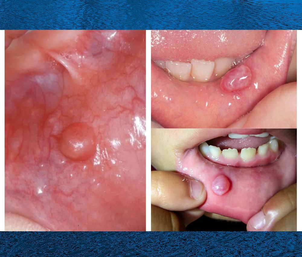
Dental Article 🔽 Oral Mucocele in Pediatric Patients: Clinical Features and Surgical Management ... Mucoceles are mucous-filled cystic lesions primarily resulting from trauma to minor salivary glands. They commonly appear in children and adolescents, particularly on the lower lip.Accurate differentiation is essential to ensure proper diagnosis, treatment planning, and recurrence prevention.
Advertisement
✅ Definition and Clinical Characteristics
➤ Mucocele
A mucocele is a mucus-filled cystic lesion arising from minor salivary glands, typically due to mechanical trauma causing ductal rupture.
Key clinical features:
▪️ Soft, fluctuant, dome-shaped swelling
▪️ Bluish or translucent appearance
▪️ Commonly located on the lower lip, buccal mucosa, or ventral tongue
▪️ Usually painless and variable in size
➤ Ranula
A ranula is a larger mucous lesion originating from the sublingual gland, located in the floor of the mouth. It represents a specific type of mucocele with deeper anatomical involvement.
Key clinical features:
▪️ Unilateral, bluish swelling in the floor of the mouth
▪️ May elevate the tongue or interfere with speech and swallowing
▪️ Can extend into the neck (plunging ranula)
📌 Recommended Article :
Dental Article 🔽 Blandin–Nuhn Mucocele: Etiology, Clinical Features, and Effective Management ... These lesions represent a subtype of oral mucoceles and are caused by extravasation of mucus following trauma or ductal disruption.✅ Etiology and Pathogenesis
Both lesions develop due to salivary flow disruption, but their mechanisms differ:
▪️ Mucocele: Most commonly caused by trauma or lip biting, leading to mucus extravasation into surrounding connective tissue.
▪️ Ranula: Typically results from ductal obstruction or rupture of the sublingual gland, with mucus accumulation in deeper anatomical planes.
The absence of an epithelial lining in most cases classifies them as pseudocysts.
📌 Recommended Article :
Dental Article 🔽 Recurrent Oral Ulcers in Children: Etiology and Management (Recurrent Aphthous Stomatitis) ... Recurrent aphthous stomatitis (RAS) is the most common cause of recurrent oral ulcers in children, characterized by painful ulcerations affecting oral mucosa without systemic disease.✅ Diagnosis
Diagnosis is primarily clinical, supported by imaging when necessary.
▪️ Mucocele: Clinical examination is usually sufficient.
▪️ Ranula: Ultrasound, CT, or MRI may be required to determine lesion extension, especially in suspected plunging ranula.
Histopathological analysis confirms diagnosis and excludes salivary gland neoplasms.
📌 Recommended Article :
Dental Article 🔽 Mouth Sores or Canker Sores? How to Tell the Difference and Heal Faster ... Mouth sores are common lesions that can appear on the oral mucosa and often cause discomfort when eating, speaking, or brushing. Among these, canker sores (aphthous ulcers) are the most frequent.✅ Treatment Options
Mucocele
▪️ Surgical excision of the lesion and associated minor salivary glands
▪️ Marsupialization in selected cases
▪️ Low recurrence when excision is complete
Ranula
▪️ Surgical removal of the sublingual gland is considered the gold standard
▪️ Simple drainage alone is associated with high recurrence rates
▪️ Plunging ranulas require combined intraoral and cervical approaches
📊 Comparative Table: Mucocele vs. Ranula – Key Clinical Differences
| Clinical Feature | Mucocele | Ranula |
|---|---|---|
| Primary gland involved | Minor salivary glands | Sublingual gland |
| Common location | Lower lip and buccal mucosa | Floor of the mouth |
| Size | Small to moderate | Often large |
| Risk of recurrence | Low after proper excision | High if sublingual gland is not removed |
| Potential complications | Minimal | Airway or swallowing interference |
Although mucocele and ranula share similar histopathological characteristics, their clinical behavior and management differ significantly. Ranulas require more aggressive treatment due to deeper glandular involvement and higher recurrence rates. Misdiagnosis or incomplete treatment may lead to repeated lesions and functional impairment.
📌 Recommended Article :
Dental Article 🔽 Congenital Syphilis: Dental Manifestations – Hutchinson Incisors and Mulberry Molars ... Among the most characteristic features are Hutchinson incisors and mulberry (Moon) molars, which reflect systemic disruption during tooth development caused by Treponema pallidum infection.🎯 Recommendations
▪️ Perform thorough clinical examination and imaging when indicated
▪️ Avoid simple drainage as definitive treatment for ranula
▪️ Submit all excised lesions for histopathological analysis
▪️ Educate patients on trauma-related risk factors
✍️ Conclusion
Mucocele and ranula are distinct salivary gland disorders that require accurate diagnosis and tailored management. Understanding their anatomical origin, clinical presentation, and evidence-based treatment options allows clinicians to reduce recurrence and optimize patient outcomes.
📚 References
✔ Neville, B. W., Damm, D. D., Allen, C. M., & Chi, A. C. (2016). Oral and maxillofacial pathology (4th ed.). Elsevier.
✔ Zhao, Y. F., Jia, Y., Chen, X. M., & Zhang, W. F. (2004). Clinical review of 580 ranulas. Oral Surgery, Oral Medicine, Oral Pathology, Oral Radiology, and Endodontology, 98(3), 281–287. https://doi.org/10.1016/j.tripleo.2004.03.006
✔ Baurmash, H. D. (2003). Mucocele and ranula. Journal of Oral and Maxillofacial Surgery, 61(3), 369–378. https://doi.org/10.1053/joms.2003.50074
📌 More Recommended Items
► What Are the Oral Manifestations in Oncology Patients? : Early and Advanced Manifestations
► What Are Fordyce Granules? Should You Be Concerned About Contagion?
► Traumatic White Lesions in the Pediatric Oral Cavity: Diagnosis, Prevention and Evidence-Based Treatment